. 2007 Jul 3:7:65.
doi: 10.1186/1471-2180-7-65.

Regions important for the adhesin activity of Moraxella catarrhalis Hag

Affiliations

Regions important for the adhesin activity of Moraxella catarrhalis Hag

Brian Bullard et al. BMC Microbiol. .

Abstract

Background: The Moraxella catarrhalis Hag protein, an Oca autotransporter adhesin, has previously been shown to be important for adherence of this respiratory tract pathogen to human middle ear and A549 lung cells.

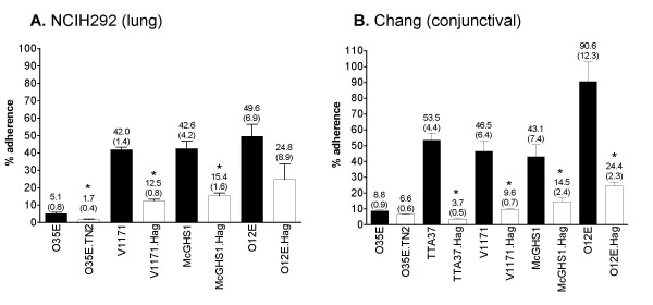
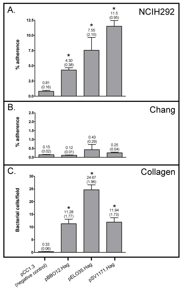
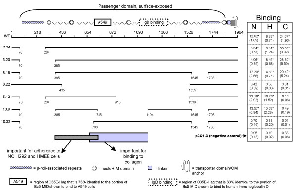
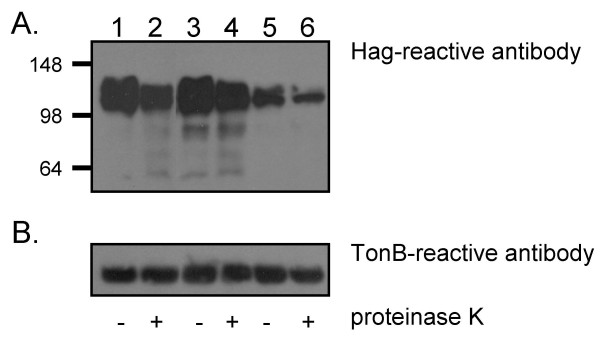
Results: The present study demonstrates that adherence of M. catarrhalis isogenic hag mutant strains to the human epithelial cell lines Chang (conjunctival) and NCIH292 (lung) is reduced by 50-93%. Furthermore, expressing Hag in a heterologous Escherichia coli background substantially increased the adherence of recombinant bacteria to NCIH292 cells and murine type IV collagen. Hag did not, however, increase the attachment of E. coli to Chang cells. These results indicate that Hag directly mediates adherence to NCIH292 lung cells and collagen, but is not sufficient to confer binding to conjunctival monolayers. Several in-frame deletions were engineered within the hag gene of M. catarrhalis strain O35E and the resulting proteins were tested for their ability to mediate binding to NCIH292 monolayers, middle ear cells, and type IV collagen. These experiments revealed that epithelial cell and collagen binding properties are separable, and that residues 385-705 of this ~2,000 amino acid protein are important for adherence to middle ear and NCIH292 cells. The region of O35E-Hag encompassing aa 706 to 1194 was also found to be required for adherence to collagen. In contrast, beta-roll repeats present in Hag, which are structural features conserved in several Oca adhesins and responsible for the adhesive properties of Yersinia enterocolitica YadA, are not important for Hag-mediated adherence.

Conclusion: Hag is a major adherence factor for human cells derived from various anatomical sites relevant to pathogenesis by M. catarrhalis and its structure-function relationships differ from those of other, closely-related autotransporter proteins.


Figures

Figure 1
Figure 1
Adherence of M. catarrhalisstrains to NCIH292 (Panel A) and Chang (Panel B) cells. Black bars correspond to WT isolates and hag isogenic mutant strains are represented by open bars. The results are expressed as the mean (± standard error) percentage of inoculated bacteria binding to monolayers. The number above each bar represents mean percentage; standard error is shown in parentheses. Asterisks indicate that the difference in adherence between a WT strain and its respective hag mutant is statistically significant.
Figure 2
Figure 2
Adherence of recombinant E. coli bacteria expressing WT Hag proteins. Panels A and B: Adherence is expressed as the mean percentage (± standard error) of inoculated bacteria binding to monolayers. Panel C: Adherence is expressed as the normalized mean number of bacteria per microscopic fields (± standard error) binding to collagen type IV coated wells. The number above each bar represents the mean; standard error is shown in parentheses. Asterisks indicate that the difference in binding between E. coli expressing O35E-Hag (i.e. pELO35.Hag), O12E-Hag (i.e. pBBO12.Hag) or V1171-Hag (i.e. pSV1171.Hag) and the negative control is statistically significant.
Figure 3
Figure 3
Selected structural features of O35E-Hag, schematic representation of in-frame deletions introduced in the protein, and adherence of recombinant E. coli bacteria expressing mutated Hag. Selected structural features are shown above in relation to their physical location within the O35E-Hag protein. Deletions within the protein are represented by gaps. Construct names are shown to the left. The columns to the right of each construct indicate the binding of E. coli expressing the mutated protein to NCIH292 cells (N), HMEE (H) or collagen (C). Adherence to NCIH292 and HMEE cells is expressed as the mean percentage (standard error shown in parentheses) of inoculated bacteria binding to monolayers. Adherence to collagen is expressed as the normalized mean number of bacteria per microscopic fields (standard error shown in parentheses) binding to collagen type IV coated wells; we estimated that 20 to 35 bacteria per microscopic field corresponds to 5–15% of input bacteria. Asterisks indicate that the difference in binding between E. coli expressing Hag proteins and the negative control is statistically significant. The negative control corresponds to E. coli carrying the plasmid control pCC1.3 and WT corresponds to recombinant bacteria harboring plasmid pELO35.Hag. The rectangles at the bottom indicate regions important for adherence to epithelial cells and collagen.
Figure 4
Figure 4
Western blot analysis of Sarkosyl-insoluble OM proteins extracted from E. coli expressing Hag proteins. OM preparations were obtained from E. coli carrying the plasmids pCC1.3 (lane 1), pELO35.Hag (lane 2), pBBHS2.24 (lane 3), pBBHS3.20 (lane 4), pBBHS8.18 (lane 5), pBBHS6.22 (lane 6), pBBHS5.12 (lane 7), pBBHS10.9 (lane 8) and pBBHS10.32 (lane 9). These preparations were resolved by SDS-PAGE, transferred to PVDF membranes and probed with antibodies against the purified recombinant protein His.Hag.CT. The figure is a composite of several western blot experiments in which OM preparations of the negative control (i.e. pCC1.3) and the positive control (i.e. pELO35.Hag) were included. Molecular weight markers are shown to the left in kDa. The numbers at the bottom of the western panel represent the predicted molecular weight of each Hag protein.
Figure 5
Figure 5
Western blot analysis of recombinant E. coli cells treated with proteinase K. E. coli carrying the plasmids pELO35.Hag (lanes 1 and 2), pBBHS2.24 (lanes 3 and 4) and pBBHS3.20 (lanes 5 and 6) were incubated for 15 min on ice in the presence (lanes 2, 4 and 6) or absence (lanes 1, 3 and 5) of proteinase K. These cells were lysed, resolved by SDS-PAGE, transferred to PVDF membranes and probed with the anti-Hag antibody 5D2 (panel A) or anti-TonB antibody 4F1 (panel B). Molecular weight markers are shown to the left in kDa.

Similar articles

Cited by

References

    1. Henderson IR, Navarro-Garcia F, Desvaux M, Fernandez RC, Ala'Aldeen D. Type V protein secretion pathway: the autotransporter story. Microbiol Mol Biol Rev. 2004;68:692–744. doi: 10.1128/MMBR.68.4.692-744.2004. - DOI - PMC - PubMed
    1. Cotter SE, Surana NK, St Geme JW., 3rd Trimeric autotransporters: a distinct subfamily of autotransporter proteins. Trends Microbiol. 2005;13:199–205. doi: 10.1016/j.tim.2005.03.004. - DOI - PubMed
    1. Hoiczyk E, Roggenkamp A, Reichenbecher M, Lupas A, Heesemann J. Structure and sequence analysis of Yersinia YadA and Moraxella UspAs reveal a novel class of adhesins. Embo J. 2000;19:5989–5999. doi: 10.1093/emboj/19.22.5989. - DOI - PMC - PubMed
    1. Cripps AW, Otczyk DC, Kyd JM. Bacterial otitis media: a vaccine preventable disease? Vaccine. 2005;23:2304–2310. doi: 10.1016/j.vaccine.2005.01.023. - DOI - PubMed
    1. Murphy TF, Brauer AL, Grant BJ, Sethi S. Moraxella catarrhalis in chronic obstructive pulmonary disease: burden of disease and immune response. Am J Respir Crit Care Med. 2005;172:195–199. doi: 10.1164/rccm.200412-1747OC. - DOI - PMC - PubMed

Publication types

LinkOut - more resources