Review
. 2007;8(6):218.
doi: 10.1186/gb-2007-8-6-218.

The calpains: modular designs and functional diversity

Affiliations
Review

The calpains: modular designs and functional diversity

Dorothy E Croall et al. Genome Biol. 2007.

Abstract

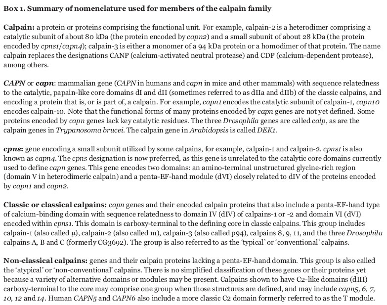
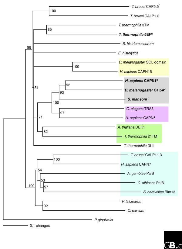
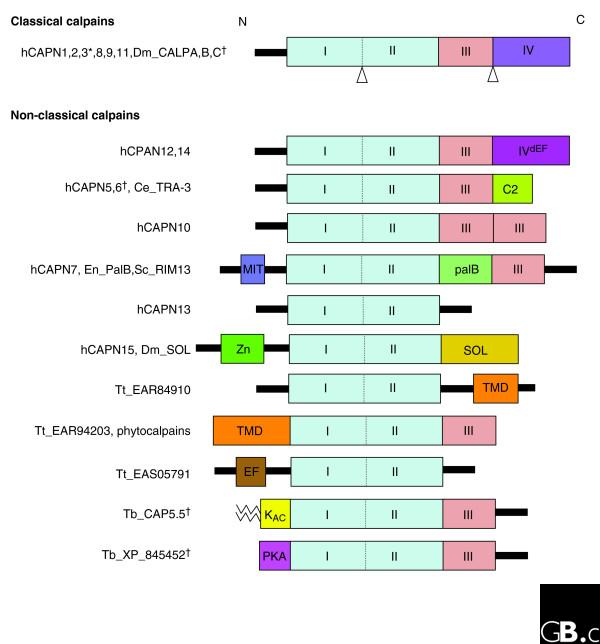
The calpain family is named for the calcium dependence of the papain-like, thiol protease activity of the well-studied ubiquitous vertebrate enzymes calpain-1 (mu-calpain) and calpain-2 (m-calpain). Proteins showing sequence relatedness to the catalytic core domains of these enzymes are included in this ancient and diverse eukaryotic protein family. Calpains are examples of highly modular organization, with several varieties of amino-terminal or carboxy-terminal modules flanking a conserved core. Acquisition of the penta-EF-hand module involved in calcium binding (and the formation of heterodimers for some calpains) seems to be a relatively late event in calpain evolution. Several alternative mechanisms for binding calcium and associating with membranes/phospholipids are found throughout the family. The gene family is expanded in mammals, trypanosomes and ciliates, with up to 26 members in Tetrahymena, for example; in striking contrast to this, only a single calpain gene is present in many other protozoa and in plants. The many isoforms of calpain and their multiple splice variants complicate the discussion and analysis of the family, and challenge researchers to ascertain the relationships between calpain gene sequences, protein isoforms and their distinct or overlapping functions. In mammals and plants it is clear that a calpain plays an essential role in development. There is increasing evidence that ubiquitous calpains participate in a variety of signal transduction pathways and function in important cellular processes of life and death. In contrast to relatively promiscuous degradative proteases, calpains cleave only a restricted set of protein substrates and use complex substrate-recognition mechanisms, involving primary and secondary structural features of target proteins. The detailed physiological significance of both proteolytically active calpains and those lacking key catalytic residues requires further study.


Figures

Box 1
Box 1
Figure 1
Figure 1
The phylogenetic relationship of calpains from diverse evolutionary groups of eukaryotes. Only the catalytic core domains (dI-II) were used to construct the tree. Multiple alignments were done with Clustal X and bootstrapped with PAUP4* (1,000 iterations). Only values greater than 50% are indicated. The tree was rooted with the calpain-related sequence from the prokaryote Porphyromonas gingivalis. A minus sign (-) indicates a nonstandard catalytic triad; species names in bold contain EF-hand motifs and the amino- or carboxy-terminal location of the motif is indicated by superscript N or C. Gray box, representative examples of classical calpains; yellow box, calpains containing a carboxy-terminal SOL domain; magenta box, calpains containing an additional carboxy-terminal C2 domain (also referred to as a Tra3 or T domain); green box, calpains containing 21 amino-terminal transmembrane domains; blue box, calpains containing a carboxy-terminal PalB-type domain. Species names: T. brucei, Trypanosoma brucei; T. thermophila, Tetrahymena thermophila; S. histriomuscorum, Sterkiella histriomuscorum (a ciliate); E. histolytica, Entamoeba histolytica; D. melanogaster, Drosophila melanogaster; S. mansoni, Schistosoma mansoni; C. elegans, Caenorhabditis elegans; H. sapiens, Homo sapiens; A. thaliana, Arabidopsis thaliana; A. gambiae, Anopheles gambiae; C. albicans, Candida albicans; S. cerevisiae, Saccharomyces cerevisiae; P. falciparum, Plasmodium falciparum; C. parvum, Cryptosporidium parvum; P. gingivalis, Porphyromonas gingivalis. Calpains listed with unpublished, nonstandard abbreviations: 3TM, three carboxy-terminal transmembrane domains; 5EF, five-EF-hand motifs; 21TM, 21 amino-terminal transmembrane domains; DI-II, domains dI-dII-only calpain without further recognizable motifs. Single calpains have been identified in organisms where only species names are given. Sequences and accession numbers are available in Additional data file 1.
Figure 2
Figure 2
A modular architecture is found in all members of the calpain protein family. All the identified human calpain genes (hCAPN) are depicted with selected examples from other species. The presence of domains dI and dII is used to define the family. Domain dIII is defined as the classical calpain C2-like domain; other C2 domains can also be present (see hCAPN5 and 6). Domain dIV is the penta-EF-hand module shared by classical calpains and their small subunit Cpns-1 (where the penta-EF-hand module is known as domain dVI). Domain dV, specific to the small subunit Cpns-1 and without known motifs, is not shown here. The black bars linking modules represent sequences without known motifs and are unique to individual calpains. *The classical calpain hCAPN3 has two insertions, indicated by Δ here. These proteins have lost key catalytic residues and are predicted to lack protease activity. Species: Dm, Drosophila melanogaster; Ce, Caenorhabiditis elegans; En, Emericella (Aspergillus) nidulans; Sc, Saccharomyces cerevisiae; Tt, Tetrahymena thermophila; Tb, Trypanosoma brucei. Domain abbreviations: C2, protein kinase C conserved region 2 (domain involved in calcium-dependent phospholipid binding); IVdEF, domain dIV with degenerate EF-hand motifs that are unlikely to bind calcium; EF, domain with EF-hand motifs distinct from domain dIV; KAC, kinetoplastid acylated domain (myristic acid and palmitic acid chains are indicated by zigzag lines); MIT, microtubule interacting and trafficking molecule domain; palB, palB-homologous domain; PKA, protein kinase A regulatory subunit domain; SOL, small optic lobe domain; TMD, transmembrane domain; Zn, zinc finger domain. The functions of some of these protein modules are not yet defined. The domain structures were assembled using SMART [79] and the peptidase database MEROPS [80].
Figure 3
Figure 3
Structures of calpain modules and calpain-2. (a) Ribbon diagram of the structure of the penta-EF-hand module (domain dVI) of Cpns-1 from pig. It is shown here as a homodimer (one chain green, one cyan). The short helical peptides (yellow and magenta) are 19-residue mimics of the conserved C peptide of the calpain inhibitor calpastatin bound to dVI in the presence of calcium (orange spheres). The structure is from PDB 1NX1 (Todd et al. [19]). (b) Ribbon diagram of the structure of the rat calpain-2 heterodimer. The catalytic core domains (dI-dII) are in light and dark blue, respectively. Catalytic residues are shown as magenta sticks (with the engineered mutation of C105S) and the arrow designates the active-site cleft between domains dI and dII. Domain dIII (brown) is C2-like. The penta-EF-hand domain dIV of the large subunit (Capn-2) is in yellow, and the similar domain dVI of the small subunit (Cpns-1) is in orange. Domain dV, the amino-terminal glycine-rich region of the small subunit, was truncated by protein engineering; in the human enzyme it is highly flexible and structurally unresolved [21]. The amino-terminal helix and linker loops are in green. The structure is from PDB 1DF0 (Hosfield et al. [20]). The dVI heterodimer in (a) is very similar to that formed between the dIV and dVI domains, and can be used to model this interaction. (c) Ribbon diagram of the structure of the calcium-bound catalytic core (domains dI-dII) of rat calpain-1 based on PDB 1TL9 (Moldoveanu et al. [26]). The bound inhibitor leupeptin is shown as gold, blue and red spheres; the magenta spheres are two calcium ions bound to hitherto unknown sites. All ribbon diagrams were generated using PyMol (DeLano Scientific, Palo Alto, CA, USA).

Similar articles

Cited by

References

    1. Gardner MJ, Bishop R, Shah T, de Villiers EP, Carlton JM, Hall N, Ren Q, Paulsen IT, Pain A, Berriman M, et al. Genome sequence of Theileria parva, a bovine pathogen that transforms lymphocytes. Science. 2005;309:134–137. doi: 10.1126/science.1110439. - DOI - PubMed
    1. Gardner MJ, Hall N, Fung E, White O, Berriman M, Hyman RW, Carlton JM, Pain A, Nelson KE, Bowman S, et al. Genome sequence of the human malaria parasite Plasmodium falciparum. Nature. 2002;419:498–511. doi: 10.1038/nature01097. - DOI - PMC - PubMed
    1. Abrahamsen MS, Templeton TJ, Enomoto S, Abrahante JE, Zhu G, Lancto CA, Deng M, Liu C, Widmer G, Tzipori S, et al. Complete genome sequence of the apicomplexan, Cryptosporidium parvum. Science. 2004;304:441–445. doi: 10.1126/science.1094786. - DOI - PubMed
    1. Loftus B, Anderson I, Davies R, Alsmark UC, Samuelson J, Amedeo P, Roncaglia P, Berriman M, Hirt RP, Mann BJ, et al. The genome of the protist parasite Entamoeba histolytica. Nature. 2005;433:865–868. doi: 10.1038/nature03291. - DOI - PubMed
    1. McArthur AG, Morrison HG, Nixon JE, Passamaneck NQ, Kim U, Hinkle G, Crocker MK, Holder ME, Farr R, Reich CI, et al. The Giardia genome project database. FEMS Microbiol Lett. 2000;189:271–273. doi: 10.1111/j.1574-6968.2000.tb09242.x. - DOI - PubMed

Publication types