Clinical Trial
. 2007 Sep;81(18):9707-17.
doi: 10.1128/JVI.00887-07. Epub 2007 Jul 3.

Human T-cell leukemia virus type 1 (HTLV-1) p12I down-modulates ICAM-1 and -2 and reduces adherence of natural killer cells, thereby protecting HTLV-1-infected primary CD4+ T cells from autologous natural killer cell-mediated cytotoxicity despite the reduction of major histocompatibility complex class I molecules on infected cells

Affiliations
Clinical Trial

Human T-cell leukemia virus type 1 (HTLV-1) p12I down-modulates ICAM-1 and -2 and reduces adherence of natural killer cells, thereby protecting HTLV-1-infected primary CD4+ T cells from autologous natural killer cell-mediated cytotoxicity despite the reduction of major histocompatibility complex class I molecules on infected cells

Prabal Banerjee et al. J Virol. 2007 Sep.

Abstract

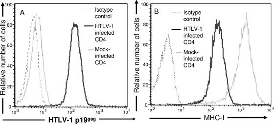
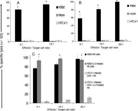
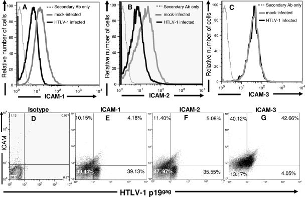
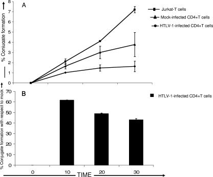
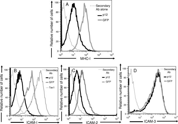
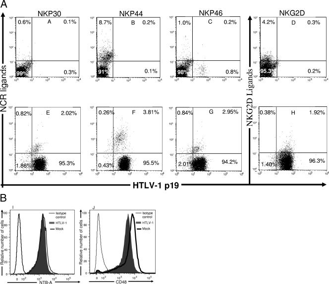
Although natural killer (NK) cell-mediated control of viral infections is well documented, very little is known about the ability of NK cells to restrain human T-cell leukemia virus type 1 (HTLV-1) infection. In the current study we show that NK cells are unable to kill HTLV-1-infected primary CD4+ T cells. Exposure of NK cells to interleukin-2 (IL-2) resulted in only a marginal increase in their ability to kill HTLV-1-infected primary CD4+ T cells. This inability of NK cells to kill HTLV-1-infected CD4+ T cells occurred despite the down-modulation of major histocompatibility complex (MHC) class I molecules, one of the ligands for the major NK cell inhibitory receptor, by HTLV-1 p12(I) on CD4+ T cells. One reason for this diminished ability of NK cells to kill HTLV-1-infected cells was the decreased ability of NK cells to adhere to HTLV-1-infected cells because of HTLV-1 p12(I)-mediated down-modulation of intercellular adhesion molecule 1 (ICAM-1) and ICAM-2. We also found that HTLV-1-infected CD4+ T cells did not express ligands for NK cell activating receptors, NCR and NKG2D, although they did express ligands for NK cell coactivating receptors, NTB-A and 2B4. Thus, despite HTLV-1-mediated down-modulation of MHC-I molecules, HTLV-1-infected primary CD4+ T cells avoids NK cell destruction by modulating ICAM expression and shunning the expression of ligands for activating receptors.


Figures

FIG. 1.
FIG. 1.
Down-modulation of MHC-I expression on primary HTLV-1-infected CD4+ T cells. HTLV-1-infected and mock-infected primary CD4+ T-cell blasts were stained with fluorochrome-conjugated anti-MHC-I antibody and then permeabilized, fixed, and stained with anti-HTLV-1 capsid antibody (p19gag) and analyzed by flow cytometry. (A) Histogram demonstrating HTLV-1 p19gag expression on 104 HTLV-1-infected (black line) or mock-infected (gray line) cells. Thin black lines represent cells stained with secondary antibody in absence of primary antibody. (B) Histogram demonstrating MHC-I expression on 104 HTLV-1-infected (black line) or mock-infected (gray line) cells. Thin black lines represent cells stained with immunoglobulin of similar isotype as MHC-I antibody.
FIG. 2.
FIG. 2.
HTLV-1-infected primary CD4+ T cells are resistant to lysis by autologous natural killer cells. HTLV-1-infected CD4+ T cells, mock-infected CD4+ T cells, and K562 cells were used as target cells in a 4-h cytotoxic assay with “resting”(A and B) or IL-2-treated (C) (1,000 U of IL-2/ml overnight) autologous NK cells as effector cells. E:T ratios of 5:1, 10:1, and 20:1 were used. All groups were performed in triplicates. Each experiment involved CD4+ T cells and NK cells from two different donors. Statistical analysis was performed by using single-tail analysis of variance and Student t tests (*, P < 0.05).
FIG. 3.
FIG. 3.
HTLV-1 infection modulates ICAM-1 and -2 expression on HTLV-1-infected primary CD4+ T cells. Mock-infected and HTLV-1-infected CD4+ T cells were indirectly stained with anti-CD54 (ICAM-1) (A and E), anti-CD102a (ICAM-2) (B and F), or anti-CD50 (ICAM-3) (C and G) antibodies. Cells were then permeabilized, fixed, and stained with anti-HTLV-1 p19 capsid antibody. The extent of ICAM-1 (A), ICAM-2 (B), and ICAM-3 (C) expression on 104 infected (thick black line) and mock-infected CD4+ T cells (thick gray line) was determined. Negative controls (dotted gray line) consisted of secondary antibody in the absence of primary antibody. (D to G) Dot plots of ICAM-1 (E), ICAM-2 (F), and ICAM-3 (G) expression versus HTLV-1 p19 capsid protein expression on 104 HTLV-1-infected CD4+ T cells. Numbers represent percentage of cells in each quadrant.
FIG. 4.
FIG. 4.
HTLV-1-infected CD4+ T cells have a decreased ability to adhere to autologous natural killer cells. (A) Heteroconjugate formation between Jurkat T cells, HTLV-1-infected CD4+ T cells, or uninfected CD4+ T cells and autologous IL-2-treated NK cells was measured at 0, 10, 20, and 30 min. Experiments were done using three separate healthy donors. (B) Percent heteroconjugate formation by HTLV-1-infected CD4+ T cells over time with respect to 100% heteroconjugate formation by mock-infected cells. All groups were evaluated in triplicate with cells from three different donors. Statistical analysis was performed by using single-tail analysis of variance and Student t tests (*, P < 0.05).
FIG. 5.
FIG. 5.
HTLV-1 p12I down-modulates MHC-I, ICAM-1, and ICAM-2 expressions on primary CD4+ T cells. Primary CD4+ T cells were transduced with lentivirus vectors encoding both GFP and HTLV-1 p12I (black line). As a control CD4+ cells were transduced with a vector expressing GFP only (gray line). Transduced CD4+ T cells were stained with PE-conjugated anti-MHC-I antibody (W6/32) (A), anti-CD54 (ICAM-1) (B), anti-CD102a (ICAM-2) (C), or anti-CD50 (ICAM-3) (D) antibody. The histogram represents gated 104 GFP-positive cells. Negative controls (gray dotted line) consisted of cells stained with secondary antibody in the absence of primary antibody.
FIG. 6.
FIG. 6.
Expression of ligands for NCR, NKG2D, 2B4, and NTB-A on HTLV-1-infected primary CD4+ T cells. Mock-infected (A to D) and HTLV-1-infected (E to H) CD4+ T cells were stained with recombinant human NKp30 and IgG Fc fusion proteins (A and E), human NKp44 and IgG Fc fusion proteins (B and F), human NKp56 and IgG Fc fusion proteins (C and G), and human NKG2D and IgG Fc fusion proteins (D and H). Cells were then permeabilized, fixed, and stained with anti-HTLV-1 p19 capsid antibody. Numbers represent the percentage of cells in each quadrant. Mock-infected and HTLV-1-infected CD4+ T cells were stained with mouse anti-human NTB-A (I) and CD48 (J) antibody. Cells were then permeabilized, fixed, and stained with anti-HTLV-1 p19 capsid antibody. The extent of CD48 and NTB-A expressed on 104 infected cells (filled gray line) and 104 mock-infected CD4+ T cells (thick black line) were determined. Negative controls (thin black line) consisted of secondary antibody in the absence of primary antibody.

Similar articles

Cited by

References

    1. Albrecht, B., N. D. Collins, M. T. Burniston, J. W. Nisbet, L. Ratner, P. L. Green, and M. D. Lairmore. 2000. Human T-lymphotropic virus type 1 open reading frame I p12I is required for efficient viral infectivity in primary lymphocytes. J. Virol. 74:9828-9835. - PMC - PubMed
    1. Arnon, T. I., M. Lev, G. Katz, Y. Chernobrov, A. Porgador, and O. Mandelboim. 2001. Recognition of viral hemagglutinins by NKp44 but not by NKp30. Eur. J. Immunol. 31:2680-2689. - PubMed
    1. Bangham, C. R., and M. Osame. 2005. Cellular immune response to HTLV-1. Oncogene 24:6035-6046. - PubMed
    1. Barber, D. F., M. Faure, and E. O. Long. 2004. LFA-1 contributes an early signal for NK cell cytotoxicity. J. Immunol. 173:3653-3659. - PubMed
    1. Barber, D. F., and E. O. Long. 2003. Coexpression of CD58 or CD48 with intercellular adhesion molecule 1 on target cells enhances adhesion of resting NK cells. J. Immunol. 170:294-299. - PubMed

Publication types

MeSH terms

Substances