. 2007 Sep;81(18):10072-80.
doi: 10.1128/JVI.00541-07. Epub 2007 Jul 3.

Regulation of hepatitis B virus replication by the phosphatidylinositol 3-kinase-akt signal transduction pathway

Affiliations

Regulation of hepatitis B virus replication by the phosphatidylinositol 3-kinase-akt signal transduction pathway

Haitao Guo et al. J Virol. 2007 Sep.

Abstract

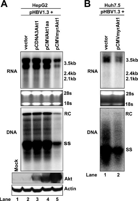
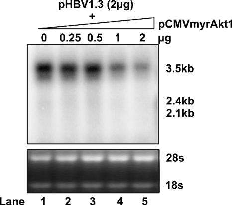
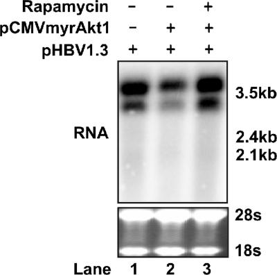
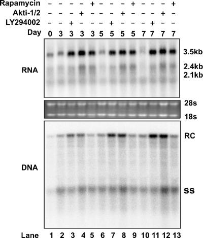
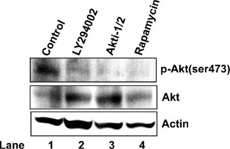
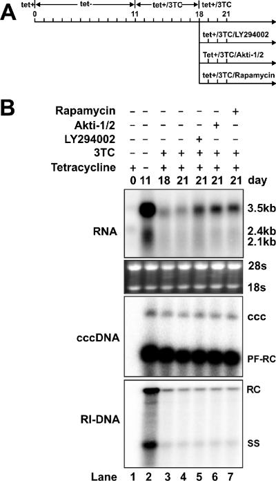
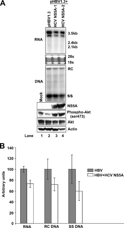
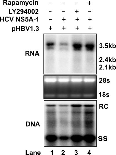
The phosphatidylinositol 3-kinase (PI3K)-protein kinase B (Akt) signaling pathway is one of the major oncogenic pathways and is activated in many types of human cancers, including hepatocellular carcinoma. It can also be activated by the hepatitis C virus (HCV) nonstructural 5A (NS5A) protein. In the present study, we set out to determine the regulatory effects of this pathway on the replication of hepatitis B virus (HBV). Our results demonstrate that the expression of a constitutively active Akt1 profoundly inhibited HBV RNA transcription and consequently reduced HBV DNA replication in HepG2 cells. This suppression of HBV gene transcription was apparently mediated by the activation of mTOR, as it was abolished by the mTOR inhibitor rapamycin. Moreover, treatment of HBV-expressing HepG2.2.15 cells with inhibitors of PI3K, Akt, and mTOR increased the transcription of 3.5-kb and 2.4-kb viral RNA as well as the replication of HBV DNA. This observation implies that the basal level activation of this pathway in HepG2 cells regulated HBV replication. Consistent with previous reports showing that the HCV NS5A protein could bind to the p85 subunit of PI3K and activate the PI3K-Akt signal transduction pathway, our results showed that expression of this protein could inhibit HBV RNA transcription and reduce HBV DNA replication in HepG2 cells. Taken together, our results suggest that the activation of the PI3K-Akt pathway during liver oncogenesis may be at least partially responsible for the elimination of HBV replication from tumor cells and may also provide an explanation for the observed suppression of HBV replication by HCV coinfection.


Figures

FIG. 1.
FIG. 1.
Effects of Akt1 activation on HBV RNA transcription and DNA replication in hepatocyte-derived cell lines. (A) A plasmid that carries pgRNA of wild-type HBV (pHBV1.3) was cotransfected with the control plasmid pUC19 (lane 2) or plasmids that express the wild-type (lane 3), dominant negative (lane 4), and constitutively active Akt1 (lane 5) forms into HepG2 cells. In these transfection experiments, 2 μg of pHBV1.3 was cotransfected with 2 μg of vector DNA or Akt1 expression plasmids into a 35-mm dish of cells. Cells were harvested at day 5 after transfection and viral RNA transcription, and DNA replication was determined by Northern (upper panel) and Southern (middle panel) blot hybridization analyses, respectively. For RNA analysis, each lane was loaded with 10 μg of total RNA. Ribosomal RNAs (28S and 18S) were presented as loading controls. The positions of HBV 3.5-kb, 2.4-kb, and 2.1-kb RNAs are indicated. For DNA analysis, HBV core DNA was probed with a genome-length minus strand-specific HBV riboprobe. The positions of relaxed circular (RC) and single-stranded (SS) DNA are indicated. Expression of Akt1 by transfected plasmids was determined by Western blot analysis, and the levels of β-actin served as loading controls (lower panel). (B) Huh7.5 cells were cotransfected with 2 μg of pHBV1.3 and 2 μg of pUC19 or pCMVmyr-Akt1. Cells were harvested at day 5 after transfection, and viral RNA transcription and DNA replication were determined by Northern (upper panel) and Southern (lower panel) blot hybridization analyses as described above.
FIG. 2.
FIG. 2.
Dose response of constitutively active Akt1 on HBV RNA transcription. The HBV DNA genome (2 μg) was cotransfected into a 35-mm dish of HepG2 cells with either 2 μg of the control vector pUC19 (lane 1) or 0.5 to 2 μg (lanes 2 to 5) of pCMVmyrAkt1. When less than 2 μg of pCMVmyrAkt1 was used for cotransfection, the control vector pUC19 was added to bring the amount of cotransfected DNA up to 2 μg. Total RNA was extracted at day 5 posttransfection and analyzed by Northern blot hybridization. rRNA served as the loading control.
FIG. 3.
FIG. 3.
Rapamycin rescues the inhibition of HBV transcription by Akt1 activation. The HBV DNA genome (2 μg) was cotransfected into HepG2 cells in a 35-mm dish with either 2 μg of the control plasmid (lane 1) or pCMVmyrAkt1 (lanes 2 and 3). Twelve hours after transfection, rapamycin was added to the culture medium to a final concentration of 10 ng/ml (lane 3); DMSO concentration in all experiment groups was normalized at 0.01%. Culture media and the inhibitor were replaced every other day, and cells were harvested at day 5 after transfection. Total RNA was extracted and analyzed by Northern blot hybridization. rRNA served as the loading control.
FIG. 4.
FIG. 4.
Effects of kinase inhibitors on HBV replication in HepG2.2.15 cells. HepG2.2.15 cells cultured in 35-mm dishes were left untreated or treated with LY294002 (10 μM), Akti-1/2 (5 μM), and rapamycin (10 ng/ml). The DMSO concentration for all experimental groups was normalized at 0.1%. Cells were harvested at days 3, 5, and 7 of treatment. Total RNA and HBV DNA replicative intermediates were extracted and analyzed by Northern blot and Southern blot hybridization, respectively.
FIG. 5.
FIG. 5.
Suppression of Akt1 activation by PI3K, Akt, and mTOR inhibitors. HepG2.2.15 cells cultured in 35-mm dishes were untreated (lane 1) or treated with LY294002 (10 μM), Akti-1/2 (5 μM), or rapamycin (10 ng/ml). The DMSO concentration in all experimental groups was normalized at 0.1%. Three days after treatment, cells were harvested, and total levels of Akt1 and Ser-473-phosphorylated Akt1 [p-Akt(ser473)] were determined by Western blot analysis as described in Materials and Methods. β-Actin served as a loading control.
FIG. 6.
FIG. 6.
The PI3K-Akt signaling pathway regulates HBV cccDNA-dependent transcription. (A) Experimental procedure. HepAD38 cells were cultured in 35-mm dishes until confluent, and tetracycline was then removed from the culture medium to induce HBV replication and cccDNA formation. After 11 days, tetracycline and lamivudine (3TC) were added to the culture medium to shut off the viral pgRNA transcription from the integrated HBV genome and to arrest viral DNA replication. Seven days later, one set of cells (lane 4) continued to be cultured with medium containing tetracycline and 3TC, while another three sets of cells were treated with 10 μM LY294002 (lane 5), 5 μM Akti-1/2 (lane 6), and 10 ng/ml rapamycin (lane 7) in the presence of tetracycline and 3TC for 3 days. The DMSO concentration in all experiment groups was normalized at 0.1%. (B) Cells were harvested at the indicated time points, and the levels of viral RNA and viral DNA replicative intermediates (RI-DNA) were determined by Northern blot and Southern blot assays, as described in Materials and Methods. As shown in the middle panel, HBV cccDNA was extracted with a modified Hirt procedure, and samples were denatured at 85°C for 5 min before loading to increase the sensitivity of cccDNA detection. Under this condition, cccDNA is not denatured, but protein-free rcDNA (PF-RC) species are denatured into single-stranded DNA (61).
FIG. 7.
FIG. 7.
HCV NS5A activates Akt1 and inhibits HBV replication. (A). The wild-type HBV genome (pHBV1.3) was cotransfected into HepG2 cells with control vector pUC19 (lane 2), wild-type HCV NS5A-expressing plasmid (HCV NS5A-1) (lane 3), or interferon-resistant HCV NS5A-expressing plasmid (HCV NS5A-2) (lane 4). In these transfection experiments, 2 μg of HBV DNA was cotransfected with 2 μg of vector DNA or HCV NS5A expression plasmid into a 35-mm dish of cells. Five days after transfection, cells were harvested, and total RNA, HBV DNA replicative intermediates, and protein were extracted and analyzed by Northern blotting, Southern blotting, and immunoblotting assays, as described in Materials and Methods. (B). HepG2 cells were cotransfected with plasmids pHBV1.3 and pUC19 or a plasmid that expresses HCV NS5A-1 and were harvested at day 4 posttransfection. Intracellular HBV RNA and DNA were assayed as described above and quantified using Bio-Rad QuantityOne software. Results from triplicate experiments are presented.
FIG. 8.
FIG. 8.
Inhibition of HBV replication by HCV NS5A can be rescued by treatment with Akt and mTOR inhibitors. The wild-type HBV genome was cotransfected into HepG2 cells with the control vector pUC19 (lane 1) or the wild-type HCV NS5A-expressing plasmid (HCV NS5A-1) (lanes 2 to 4). In these transfection experiments, 2 μg of HBV DNA was cotransfected with 2 μg of vector DNA or HCV NS5A expression plasmid into a 35-mm dish of cells. Twelve hours after transfection, LY294002 (lane 3) or rapamycin (lane 4) was added to the culture medium at concentrations described above. Cells were harvested at day 5 after transfection. The viral RNA and DNA levels were determined by Northern blotting and Southern blotting assays as described in Materials and Methods.

References

    1. Acs, G., M. A. Sells, R. H. Purcell, P. Price, R. Engle, M. Shapiro, and H. Popper. 1987. Hepatitis B virus produced by transfected Hep G2 cells causes hepatitis in chimpanzees. Proc. Natl. Acad. Sci. USA 84:4641-4644. - PMC - PubMed
    1. Barnett, S. F., D. Defeo-Jones, S. Fu, P. J. Hancock, K. M. Haskell, R. E. Jones, J. A. Kahana, A. M. Kral, K. Leander, L. L. Lee, J. Malinowski, E. M. McAvoy, D. D. Nahas, R. G. Robinson, and H. E. Huber. 2005. Identification and characterization of pleckstrin-homology-domain-dependent and isoenzyme-specific Akt inhibitors. Biochem. J. 385:399-408. - PMC - PubMed
    1. Beckel-Mitchener, A., and J. Summers. 1997. A novel transcriptional element in circular DNA monomers of the duck hepatitis B virus. J. Virol. 71:7917-7922. - PMC - PubMed
    1. Blight, K. J., J. A. McKeating, and C. M. Rice. 2002. Highly permissive cell lines for subgenomic and genomic hepatitis C virus RNA replication. J. Virol. 76:13001-13014. - PMC - PubMed
    1. Bouchard, M. J., R. J. Puro, L. Wang, and R. J. Schneider. 2003. Activation and inhibition of cellular calcium and tyrosine kinase signaling pathways identify targets of the HBx protein involved in hepatitis B virus replication. J. Virol. 77:7713-7719. - PMC - PubMed

Publication types

MeSH terms

LinkOut - more resources