Comparative Study
. 2007 Jul 10;104(28):11670-5.
doi: 10.1073/pnas.0704692104. Epub 2007 Jul 3.

Laboratory evolution of one disulfide isomerase to resemble another

Affiliations
Comparative Study

Laboratory evolution of one disulfide isomerase to resemble another

Annie Hiniker et al. Proc Natl Acad Sci U S A. .

Abstract

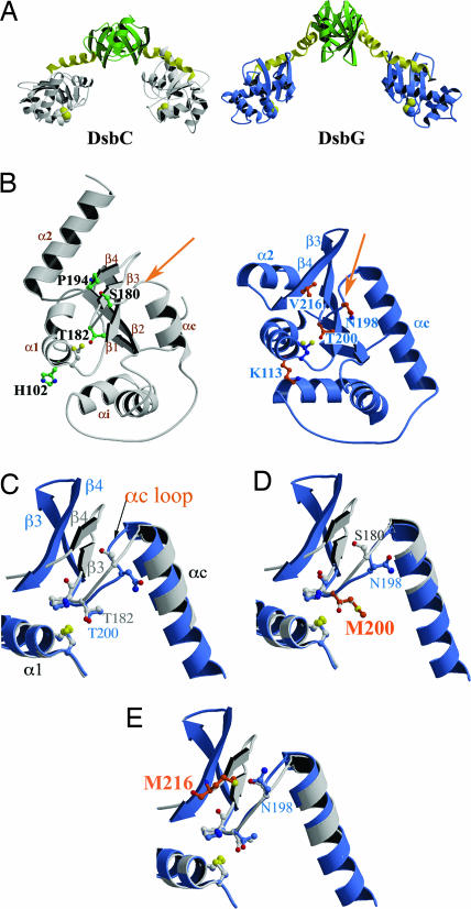
It is often difficult to determine which of the sequence and structural differences between divergent members of multigene families are functionally important. Here we use a laboratory evolution approach to determine functionally important structural differences between two distantly related disulfide isomerases, DsbC and DsbG from Escherichia coli. Surprisingly, we found single amino acid substitutions in DsbG that were able to complement dsbC in vivo and have more DsbC-like isomerase activity in vitro. Crystal structures of the three strongest point mutants, DsbG K113E, DsbG V216M, and DsbG T200M, reveal changes in highly surface-exposed regions that cause DsbG to more closely resemble the distantly related DsbC. In this case, laboratory evolution appears to have taken a direct route to allow one protein family member to complement another, with single substitutions apparently bypassing much of the need for multiple changes that took place over approximately 0.5 billion years of evolution. Our findings suggest that, for these two proteins at least, regions important in determining functional differences may represent only a tiny fraction of the overall protein structure.


Conflict of interest statement

The authors declare no conflict of interest.

Figures

Fig. 1.
Fig. 1.
Copper-resistant phenotype of four DsbG mutants rescuing copper resistance of a dsbC-null strain (for complete list of mutants, see SI Table 1). Wt, wild-type.
Fig. 2.
Fig. 2.
Isomerase activity of DsbG mutants is enhanced. Disulfide-scrambled hirudin was incubated in the absence or in the presence of stoichiometric quantities of wild-type DsbC, DsbG, or the DsbG mutants and allowed to refold at 25°C for 18 h. Samples were acid trapped, and folding intermediates were separated by reversed-phase HPLC. The dotted lines labeled R and N denote the retention times of fully reduced and native hirudin, respectively.
Fig. 3.
Fig. 3.
Comparison of the structures of DsbC, DsbG, and DsbC-like DsbG mutants identifies key regions functionally distinguishing the two proteins. Active-site cysteines are shown in yellow in all parts of the figure. (A) The structures of DsbC and DsbG show that they are both V-shaped homodimers with a thioredoxin-like domain (gray and blue, respectively) connected by a linker helix (yellow) to an N-terminal dimerization domain (green). (B) Comparison of the thioredoxin domains of DsbC (left, gray) and DsbG (right, blue). The αc loop (indicated by an arrow) links the connecting helix (αc) with strand β3. This loop has very different conformations in DsbC and DsbG. The residues mutated in DsbG that allowed it to gain DsbC-like properties are indicated (K113, N198, T200, and V216) and the corresponding residues in DsbC are also shown (H102, S180, T182, and P194, respectively). Two of these four residues are located on the αc loop (N198 and T200). (C) Superposition of wild-type DsbC (gray) and wild-type DsbG (blue), showing that they have very different αc loop conformations. (D) Superposition of wild-type DsbC (gray) and DsbG T200M (blue, mutated residue in orange; this work, Protein Data Bank ID code 2H0G), showing that the αc loop conformation of DsbG T200M is more like that of DsbC. (E) Superposition of wild-type DsbC (gray) and DsbG V216M (blue, mutated residue in orange; PDB ID code 2H0I), showing that the αc loop conformation of DsbG V216M is more like that of DsbC.
Fig. 4.
Fig. 4.
Electrostatic surface of a portion of wild-type DsbC, wild-type DsbG, and variants of DsbG identifies a key difference between the two proteins. Positive and negative electrostatic potentials are shown in blue and red, respectively (saturation at 18 kT/e) for each of the proteins. Electrostatic surface representations were generated by using GRASP (28). These surfaces are derived from the crystal structures of wild-type DsbC (Protein Data Bank ID code 1EEJ), wild-type DsbG [PDB ID code 1V57 (8)], DsbG K113E (this work, PDB ID code 2H0H), and DsbG V216M (this work, PDB ID code 2H0I), and models of DsbG K113D, DsbG K113N, and DsbG K113W.

References

    1. Bartel DP, Unrau PJ. Trends Cell Biol. 1999;9:M9–M13. - PubMed
    1. Yuan L, Kurek I, English J, Keenan R. Microbiol Mol Biol Rev. 2005;69:373–392. - PMC - PubMed
    1. Rothman SC, Kirsch JF. J Mol Biol. 2003;327:593–608. - PubMed
    1. Jurgens C, Strom A, Wegener D, Hettwer S, Wilmanns M, Sterner R. Proc Natl Acad Sci USA. 2000;97:9925–9930. - PMC - PubMed
    1. Griswold KE, Aiyappan NS, Iverson BL, Georgiou G. J Mol Biol. 2006;364:400–410. - PMC - PubMed

Publication types

MeSH terms

LinkOut - more resources