. 2007 Jul 17;104(29):11939-44.
doi: 10.1073/pnas.0704057104. Epub 2007 Jul 3.

Structural consequences of disease-causing mutations in the ATRX-DNMT3-DNMT3L (ADD) domain of the chromatin-associated protein ATRX

Affiliations

Structural consequences of disease-causing mutations in the ATRX-DNMT3-DNMT3L (ADD) domain of the chromatin-associated protein ATRX

Anthony Argentaro et al. Proc Natl Acad Sci U S A. .

Abstract

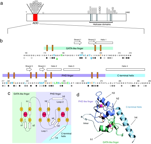
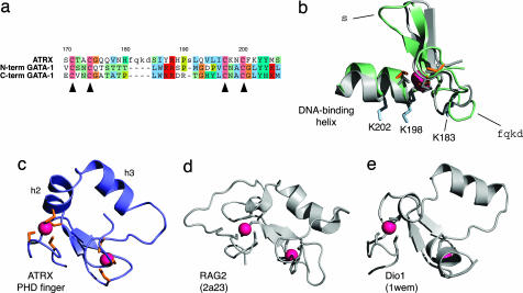
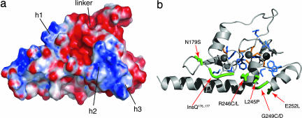
The chromatin-associated protein ATRX was originally identified because mutations in the ATRX gene cause a severe form of syndromal X-linked mental retardation associated with alpha-thalassemia. Half of all of the disease-associated missense mutations cluster in a cysteine-rich region in the N terminus of ATRX. This region was named the ATRX-DNMT3-DNMT3L (ADD) domain, based on sequence homology with a family of DNA methyltransferases. Here, we report the solution structure of the ADD domain of ATRX, which consists of an N-terminal GATA-like zinc finger, a plant homeodomain finger, and a long C-terminal alpha-helix that pack together to form a single globular domain. Interestingly, the alpha-helix of the GATA-like finger is exposed and highly basic, suggesting a DNA-binding function for ATRX. The disease-causing mutations fall into two groups: the majority affect buried residues and hence affect the structural integrity of the ADD domain; another group affects a cluster of surface residues, and these are likely to perturb a potential protein interaction site. The effects of individual point mutations on the folding state and stability of the ADD domain correlate well with the levels of mutant ATRX protein in patients, providing insights into the molecular pathophysiology of ATR-X syndrome.


Conflict of interest statement

The authors declare no conflict of interest.

Figures

Fig. 1.
Fig. 1.
ATRX protein sequence, structure, and disease-associated mutations. (a) The locations of the highly conserved N-terminal cysteine-rich domain and the C-terminal helicase-like domain are shown. The positions of missense mutations are indicated with circles and the number of times (>1) the mutation has been identified in unrelated individuals is indicated within relevant circles. All of the circles drawn between the oblique lines above the bar refer to mutations within the ADD domain. (b) Locations of mutations and secondary structural elements in the ADD domain. The N-terminal GATA-like zinc finger is indicated by a light green bar, the PHD finger by a mauve bar, and the C-terminal extension by a light blue bar. The conserved cysteine residues are marked as orange vertical bars. Missense mutations are highlighted in green (surface), blue (buried), and orange (cysteines); the insertion mutation is highlighted by an upward green arrow and the deletion by a downward blue arrow. Residues where there is homology across the whole family of ADD domain sequences (ATRX, DNMT3A, DNMT3B, and DNMT3L) are marked with filled circles (absolute conservation), gray circles (strong conservation), and open circles (weak conservation); for the full alignment, see SI Fig. 5. (c) Schematic showing the zinc-binding topology and secondary structure elements of the ADD domain, color scheme as for b. β-Strands are labeled s1–s4 and helices h1–h4. The zinc binding within the PHD finger has the “cross-braced” topology characteristic of such domains, with each zinc coordinated by a noncontiguous set of ligands. (d) Ribbon representation of the NMR structure of the ADD domain (lowest energy structure from the accepted ensemble of 32) of ATRX. The GATA-like finger is shown in green, the PHD finger in mauve, and the C-terminal helix in blue. Linker and unstructured regions are shown in gray, zinc atoms in pink, and side chains of the zinc coordinating cysteines in orange.
Fig. 2.
Fig. 2.
Sequence alignment and structural comparisons of the ADD domain with other GATA and PHD fingers. (a) Structure-based sequence alignment of the GATA-like zinc finger of ATRX with the N- and C-terminal zinc fingers of GATA-1. Residues considered as structurally equivalent are shown in uppercase, structurally dissimilar residues are shown in lowercase, and background colors follow the ClustalX scheme. Numbering is based on the ATRX sequence, and the positions of the metal-binding cysteines are indicated with triangles below the alignment. (b) Structural superposition of the GATA-like finger of the ADD domain of ATRX (color scheme as in Fig. 1 b–d) with the C-terminal zinc finger of GATA-1 (shown in gray, except for the zinc, which is red). The position in the structure of the single-residue insertion in the ADD domain relative to GATA-1 is indicated as “s” and that of the four-residue insertion is indicated as “fqkd.” The helix used for DNA binding by the C-terminal finger of GATA-1 and its analogue in the ADD domain are indicated, and the basic residues that the ADD domain might use for DNA binding (see text) are shown as side chains in blue and labeled with their sequence positions. The superposition was made by fitting the N, Cα, and C′ atoms of residues 167–180, 185–190, and 192–207 of ATRX to the corresponding residues of the C-terminal finger of GATA-1, based on the alignment in a. (c) PHD finger from the ADD domain of ATRX (color scheme as in Fig. 1 b–d). (d) Structure of the PHD finger from V(D)J recombination activating protein 2 (RAG2, Protein Data Bank ID code 2a23) (28), shown in the same orientation as in c. (e) Structure of the PHD finger from death-inducer obliterator-1 (Dio1, Protein Data Bank ID code 1wem), shown in the same orientation as in c. These PHD fingers were chosen for comparison, because they have the closest structural similarity to the PHD finger of ATRX in terms of the position and orientation of helical elements. The correct orientation in c–e was achieved by superposing the positions of the corresponding zinc-binding atoms of the eight zinc-binding residues in each protein.
Fig. 3.
Fig. 3.
Electrostatic potential and location of mutations in the structure of the ADD domain. (a) Surface electrostatic potential of the ADD domain, shown in the same orientation as the ribbon view in b. The helix in the GATA-like finger (h1) is solvent-exposed and basic, and the two helices within loop 2 of the PHD finger (h2 and h3) form another basic patch. The linker between the GATA-like and PHD fingers is highly acidic. (b) Ribbon structure of the ADD domain showing the locations of mutations found in patients with ATR-X syndrome. Mutations are classified as surface (green), buried (blue), or cysteine (orange) and are represented by using their side chains, except for the glycine mutation G249C/D and the glutamine insertion, which are represented by thickening the backbone. The surface mutations are individually labeled.
Fig. 4.
Fig. 4.
ATRX in vivo expression in EBV-transformed patient lymphocytes. (a) ATRX mRNA levels of patient mutations and normal controls as determined by quantitative RT-PCR. Patients are grouped according to the nature of their underlying mutation: cysteine mutations are orange, buried mutations are blue, and surface mutations are green. Values for normal individuals are represented by black circles. For each case, the ATRX mRNA level is expressed as the percentage of the average for 18 normal control individuals. (b) ATRX protein levels of patients and normal controls. Cases are grouped as in a. ATRX protein levels are expressed as a percentage of the average value for seven normal control individuals. (c) Representative Western blots showing ATRX protein levels (including loading control). Lane 1 represents the ATRX protein level for a cysteine mutation, lanes 2–4 are buried mutations, lanes 5–7 are surface mutations, and lane 8 is a normal control.

References

    1. Weatherall DJ, Higgs DR, Bunch C, Old JM, Hunt DM, Pressley L, Clegg JB, Bethlenfalvay NC, Sjolin S, Koler RD, et al. N Engl J Med. 1981;305:607–612. - PubMed
    1. Gibbons RJ, Picketts DJ, Villard L, Higgs DR. Cell. 1995;80:837–845. - PubMed
    1. Garrick D, Sharpe JA, Arkell R, Dobbie L, Smith AJH, Wood WG, Higgs DR, Gibbons RJ. PLoS Genet. 2006;2:438–450. - PMC - PubMed
    1. Gibbons RJ, Pellagatti A, Garrick D, Wood WG, Malik N, Ayyub H, Langford C, Boultwood J, Wainscoat JS, Higgs DR. Nat Genet. 2003;34:446–449. - PubMed
    1. McDowell TL, Gibbons RJ, Sutherland H, O'Rourke DM, Bickmore WA, Pombo A, Turley H, Gatter K, Picketts DJ, Buckle VJ, et al. Proc Natl Acad Sci USA. 1999;96:13983–13988. - PMC - PubMed

MeSH terms

Associated data