. 2007 Jul 10;104(28):11832-7.
doi: 10.1073/pnas.0702176104. Epub 2007 Jul 3.

An NQO1- and PARP-1-mediated cell death pathway induced in non-small-cell lung cancer cells by beta-lapachone

Affiliations

An NQO1- and PARP-1-mediated cell death pathway induced in non-small-cell lung cancer cells by beta-lapachone

Erik A Bey et al. Proc Natl Acad Sci U S A. .

Abstract

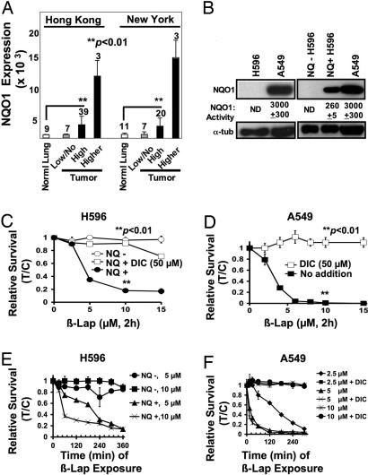
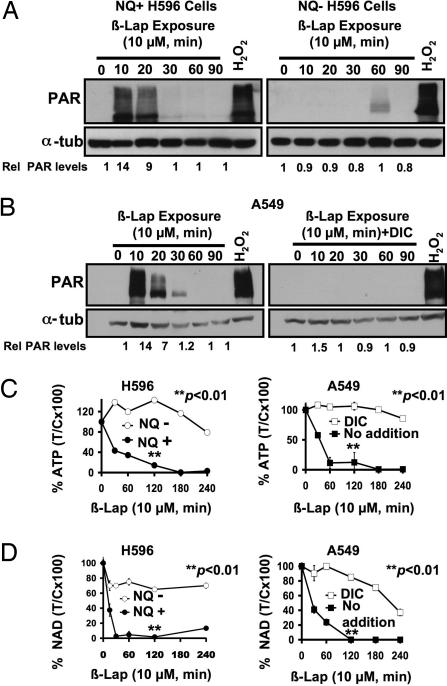
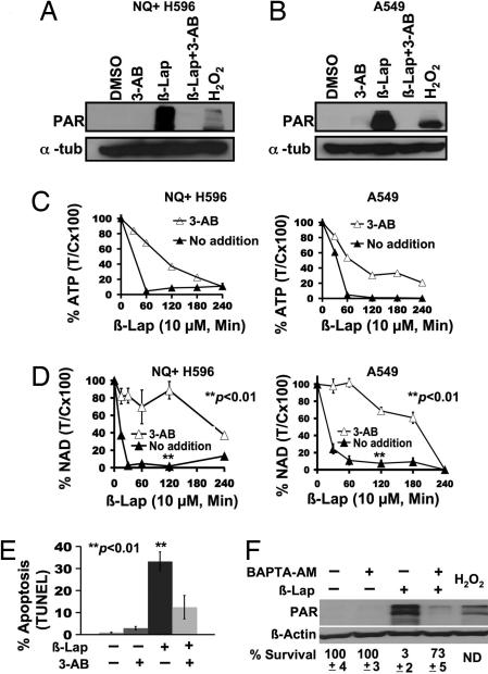
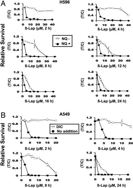
Lung cancer is the number one cause of cancer-related deaths in the world. Patients treated with current chemotherapies for non-small-cell lung cancers (NSCLCs) have a survival rate of approximately 15% after 5 years. Novel approaches are needed to treat this disease. We show elevated NAD(P)H:quinone oxidoreductase-1 (NQO1) levels in tumors from NSCLC patients. beta-Lapachone, an effective chemotherapeutic and radiosensitizing agent, selectively killed NSCLC cells that expressed high levels of NQO1. Isogenic H596 NSCLC cells that lacked or expressed NQO1 along with A549 NSCLC cells treated with or without dicoumarol, were used to elucidate the mechanism of action and optimal therapeutic window of beta-lapachone. NSCLC cells were killed in an NQO1-dependent manner by beta-lapachone (LD50, approximately 4 microM) with a minimum 2-h exposure. Kinetically, beta-lapachone-induced cell death was characterized by the following: (i) dramatic reactive oxygen species (ROS) formation, eliciting extensive DNA damage; (ii) hyperactivation of poly(ADP-ribose)polymerase-1 (PARP-1); (iii) depletion of NAD+/ATP levels; and (iv) proteolytic cleavage of p53/PARP-1, indicating mu-calpain activation and apoptosis. Beta-lapachone-induced PARP-1 hyperactivation, nucleotide depletion, and apoptosis were blocked by 3-aminobenzamide, a PARP-1 inhibitor, and 1,2-bis(2-aminophenoxy)ethane-N,N,N',N'-tetraacetic acid acetoxymethyl ester (BAPTA-AM), a Ca2+ chelator. NQO1- cells (H596, IMR-90) or dicoumarol-exposed NQO1+ A549 cells were resistant (LD50, >40 microM) to ROS formation and all cytotoxic effects of beta-lapachone. Our data indicate that the most efficacious strategy using beta-lapachone in chemotherapy was to deliver the drug in short pulses, greatly reducing cytotoxicity to NQO1- "normal" cells. beta-Lapachone killed cells in a tumorselective manner and is indicated for use against NQO1+ NSCLC cancers.


Conflict of interest statement

The authors declare no conflict of interest.

Figures

Fig. 1.
Fig. 1.
NQO1 overexpression in NSCLC confers susceptibility to β-lap. (A) DNA microarray array data of NSCLC patient samples from New York and Hong Kong tissue banks were analyzed for NQO1 mRNA levels as described in Materials and Methods. Numbers above bars indicate the number of patients analyzed in each category. Associated normal tissue from patient samples was also evaluated but was not always available. (B) NQO1 protein levels and enzyme activities (nanomoles per minute per microgram) in H596 and A549 NSCLC cells, vector-alone H596 (NQ H596) cells, and in H596 cells stably expressing NQO1 (NQ+ H596). Equivalent loading was monitored by using α-tubulin (α-tub). NQO1 activity <1.0 nmol·min−1·μg−1 was nondetectable (ND). (C) Long-term relative survival assays of NQ H596 (○) and NQ+ H596 (●) cells treated for 2 h with β-lap or β-lap plus DIC (□) at the indicated doses. Shown are means ± SE for three independent experiments performed in triplicate. (D) A549 cells were treated as described for C, with β-lap (■) with or without 50 μM DIC (□). (E) NQ+ H596 and NQ H596 cells were exposed to doses of β-lap, and drugs were removed at various times up to 360 min to determine minimum exposure times required for lethality. (F) A549 cells were treated with various β-lap doses, with or without 50 μM DIC, and long-term relative survival assays were performed. Results (means ± SE) in C–F are representative of experiments performed at least three times. In C and D, Student's t tests were performed. ∗∗, P < 0.01 vs. NQ cells treated with β-lap alone (C) and A549 cells exposed to β-lap plus DIC (D).
Fig. 2.
Fig. 2.
β-Lap induces oxidative stress and DNA damage in NQO1+ H596 and A549 cells. (A) NQ+ H596 (Left), NQ H596 (Left), and A549 cells (Right) were treated with 10 μM β-lap with or without 50 μM DIC. ROS formation was monitored at the indicated times by DCF staining as described in Materials and Methods. Cells were also treated with 2 mM H2O2 or 10 μM antimycin A for 20 min as positive controls (data not shown). Results shown are means ± SE for three or more experiments. ∗∗, P < 0.01 vs. NQ H596 (Left) and A549 cells treated with β-lap plus DIC (Right). (B) NQ+ H596, NQ H596, and A549 cells were treated with the indicated doses of β-lap with or without 50 μM DIC for 20 min, and ROS formation was measured as in A. (C) NQ H596, NQ+ H596, and A549 cells were exposed to β-lap with or without 50 μM DIC and assessed for DNA damage by using comet assays as described in Materials and Methods. Cells were also exposed to 2 mM H2O2 for 2 h as positive controls. Comet tail lengths of NQO1 and NQO1+ H596 cells were measured by using Image J software (a.u., arbitrary unit). Shown are representative micrographs of experiments performed at least three times. Graphed are means ± SE from three experiments. Student's t tests were performed. ∗∗, P < 0.001 vs. NQ H596 treated with β-lap alone.
Fig. 3.
Fig. 3.
β-Lap-induced PARP-1 hyperactivation causes NQO1-dependent nucleotide depletion. (A and B) Western blot analyses of PAR formation in NQO1+ and NQO1 H596 cells treated with or without β-lap (10 μM) (A) or A549 cells exposed to β-lap (10 μM) (B Left) with or without 50 μM DIC (B Right) and harvested at the indicated times. PAR formation was detected as described in Materials and Methods. (C) NQ+ H596, NQ H596, and A549 cells were mock-treated or exposed to β-lap (10 μM), harvested at the indicated times, and analyzed for ATP level changes as in Materials and Methods. For A549 cells, DIC (50 μM) was added with β-lap to examine the effects of NQO1 inhibition. (D) NQ+ H596, NQ H596, and A549 cells were treated with β-lap (10 μM) with or without DIC (40 μM), and changes in intracellular NAD+ levels were analyzed at indicated times as described in Materials and Methods. For C and D, results shown are means ± SE for experiments performed three or more times. Student's t tests were performed. ∗∗, P < 0.01 vs. NQ H596 cells treated with β-lap alone and A549 cells treated with β-lap plus DIC.
Fig. 4.
Fig. 4.
Inhibition of PARP-1 hyperactivation delays NQO1-dependent nucleotide pool depletion in NSCLC cells after β-lap. (A and B) NQ+ H596 or A549 cells were pretreated with or without 25 mM 3-AB or mock-treated for 2 h and then exposed to β-lap with or without 3-AB for 20 min. (C and D) NQ+ H596 and A549 cells were treated with β-lap with or without 3-AB as in A, harvested at the indicated times, and analyzed for changes in nucleotide (NAD+ and ATP) pool levels as described in Materials and Methods. Results were graphed as means ± SE of experiments performed three times. Student's t tests were performed. ∗∗, P < 0.01 vs. NQ H596 cells treated with β-lap alone; A549 cells treated with β-lap plus DIC. (E) A549 cells were mock-treated or pretreated with 3-AB as described (19) and then exposed to β-lap (10 μM, 2 h) and harvested 48 h later. Cells were assessed for apoptosis by TUNEL assays. Student's t tests were performed. ∗∗, P < 0.01 vs. A549 cells treated with β-lap plus 3-AB. (F) Western blot of A549 cells pretreated with 5 μM BAPTA-AM as described in Materials and Methods or mock-treated and then exposed to β-lap (6 μM, 10 min). Samples were then harvested for PAR formation. Survival data are expressed as %T/C. The percentage of NAD+ loss was also monitored as a surrogate for PARP-1 hyperactivation in cells pretreated with 5 μM BAPTA-AM or mock-treated and then exposed to β-lap (6 μM, 1 h). BAPTA-AM-pretreated, β-lap-exposed cells retained NAD+ (87% control), whereas β-lap-treated NQO1+ A549 cells lost significant NAD+ levels (43%) measured at 1 h after treatment (see SI Fig. 7B). ND, not determined.
Fig. 5.
Fig. 5.
Defining the therapeutic window of β-lap. (A) Long-term relative survival assays of NQ+ and NQ H596 cells treated with β-lap at the doses (micromolar) and times (hours) indicated. After various exposures, relative survival was assessed. (B) Relative survival assays of A549 cells treated with various concentrations of β-lap with or without 50 μM DIC for 2–24 h. Data are graphed as in A, except that β-lap alone vs. β-lap plus DIC were graphed. ○, NQO1 cells treated with or without various doses and times of β-lap. ● or ■, NQO1+ cells treated in the same manner. □, NQO1+ cells treated with β-lap plus 50 μM DIC to inhibit NQO1. Addition of 50 μM DIC to β-lap-treated NQO1 cells had no additional effect. The graphs show the means ± SE of three independent experiments performed in triplicate.

References

    1. Jemal A, Siegel R, Ward E, Murray T, Xu J, Smigal C, Thun MJ. CA Cancer J Clin. 2006;56:106–130. - PubMed
    1. Spira A, Ettinger DS. N Engl J Med. 2004;350:379–392. - PubMed
    1. Kim MY, Zhang T, Kraus WL. Genes Dev. 2005;19:1951–1967. - PubMed
    1. Zong WX, Ditsworth D, Bauer DE, Wang ZQ, Thompson CB. Genes Dev. 2004;18:1272–1282. - PMC - PubMed
    1. Pink JJ, Wuerzberger-Davis S, Tagliarino C, Planchon SM, Yang X, Froelich CJ, Boothman DA. Exp Cell Res. 2000;255:144–155. - PubMed

Publication types

MeSH terms