. 2007 Jul 17;104(29):11969-74.
doi: 10.1073/pnas.0701979104. Epub 2007 Jul 3.

Identifying important structural characteristics of arsenic resistance proteins by using designed three-stranded coiled coils

Affiliations

Identifying important structural characteristics of arsenic resistance proteins by using designed three-stranded coiled coils

Debra S Touw et al. Proc Natl Acad Sci U S A. .

Abstract

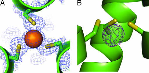
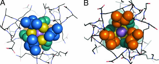
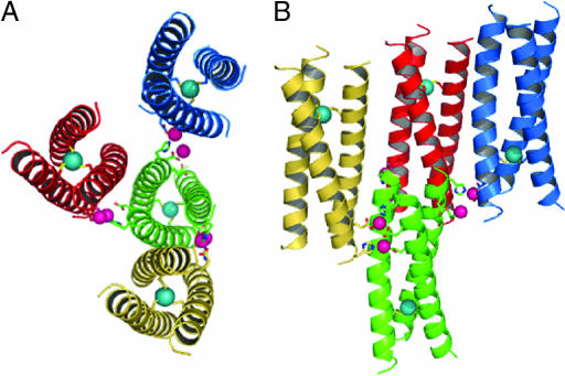
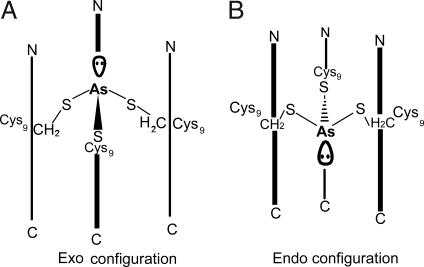
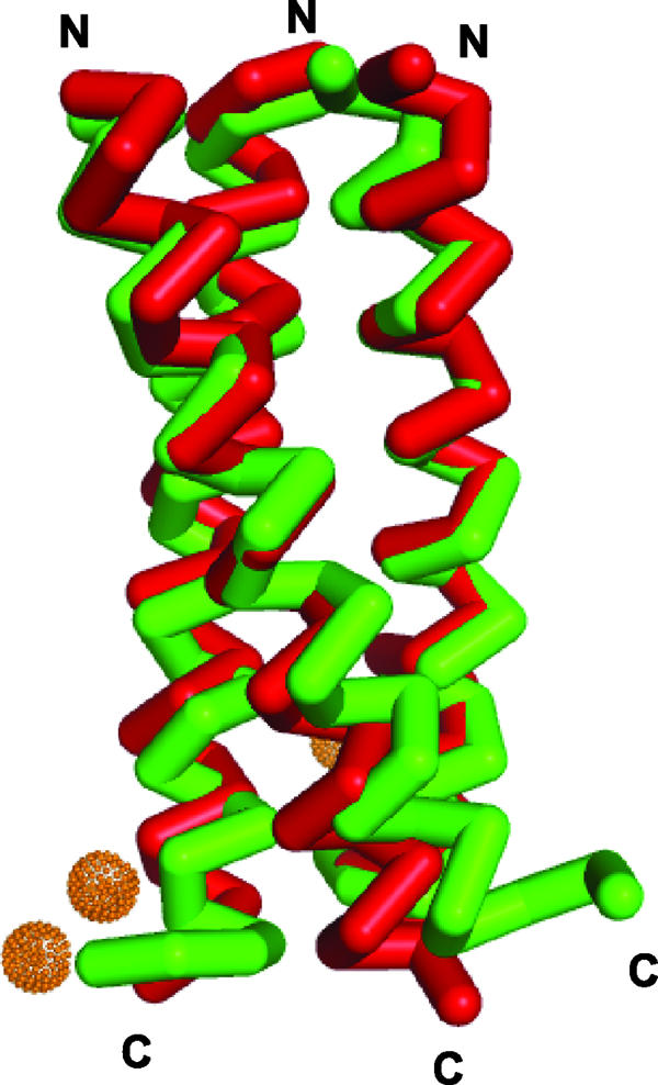
Arsenic, a contaminant of water supplies worldwide, is one of the most toxic inorganic ions. Despite arsenic's health impact, there is relatively little structural detail known about its interactions with proteins. Bacteria such as Escherichia coli have evolved arsenic resistance using the Ars operon that is regulated by ArsR, a repressor protein that dissociates from DNA when As(III) binds. This protein undergoes a critical conformational change upon binding As(III) with three cysteine residues. Unfortunately, structures of ArsR with or without As(III) have not been reported. Alternatively, de novo designed peptides can bind As(III) in an endo configuration within a thiolate-rich environment consistent with that proposed for both ArsR and ArsD. We report the structure of the As(III) complex of Coil Ser L9C to a 1.8-A resolution, providing x-ray characterization of As(III) in a Tris thiolate protein environment and allowing a structural basis by which to understand arsenated ArsR.


Conflict of interest statement

The authors declare no conflict of interest.

Figures

Fig. 1.
Fig. 1.
A top-down view from the N terminus of the coiled coil and a side view of the trigonal thiolate As(III)-binding site illustrate its pyramidal coordination in the interior of the three-stranded coiled coil. (A) Top-down view of the As(III) ion coordinated to the three cysteine residues in position 9 of the heptad repeat with 2FoFc electron density contoured at 1.5 σ overlaid. (B) Side view of the metal-binding site with an FoFc omit map contoured at 5 σ demonstrating the endo coordination of the As(III) ion 1.3 Å below the plane of the Cys Sγ-atoms and on an equal level with the β-methylene protons.
Fig. 2.
Fig. 2.
The packing of the leucine layers above and below the As(III) ion, shown as a purple sphere, is illustrated. (A) Top-down view of the leucine 5 layer (blue). (B) Leu 12 layer (orange) looking up the helical axis from the C terminus; this shows the orientation of the d residues in this layer toward the helical interface. This illustrates that if a metal that prefers tetrahedral coordination such as Cd(II) or Zn(II) were coordinated to the cysteines in the same manner as As(III), the fourth exogenous ligand (e.g., water) would coordinate on the C-terminal side of the metal ion.
Fig. 3.
Fig. 3.
The zinc-mediated packing of the coiled coils with Zn(II) and As(III) ions is shown in pink and cyan, respectively. (A) Bottom-up view of a central coiled coil (green) with the six Zn(II) ions that are coordinated by side chains at the C-terminal end. (B) Side view of the trimeric structure of As(CSL9C)3 with a different perspective of the Zn(II) coordination to the exterior residues of the coiled coils. In both panels, only two of eight symmetry related Zn(II) ions are included for clarity.
Fig. 4.
Fig. 4.
Scheme showing the two possible orientations of As(III) within a three-stranded coiled coil. (A) In the exo configuration, the As(III) ion would be located above both Sγ atoms and β-methylene protons of the cysteine side chains, and the lone pair would point toward the N terminus. (B) In the endo configuration, which is observed in the structure of As(CSL9C)3, the As(III) ion would be located below the Sγ atoms and approximately level with the β-methylene protons of the cysteine side chains. The lone pair would then be directed toward the C terminus.
Fig. 5.
Fig. 5.
The overlay of Coil VaLd, shown in green (PDB entry 1COI), a related parallel three-stranded coiled coil with As(CSL9C)3 (red) demonstrates their structural similarity and highlights their divergence at the C and N termini where the Zn(II) ions hold CSL9C in a more helical conformation than VaLd.

References

    1. Shi W, Dong J, Scott RA, Ksenzenko MY, Rosen BP. J Biol Chem. 1996;271:9291–9297. - PubMed
    1. Zhou T, Radaev S, Rosen BP, Gatti DL. EMBO J. 2000;19:4838–4845. - PMC - PubMed
    1. Martin P, DeMel S, Shi J, Gladysheva T, Gatti DL, Rosen BP, Edwards BFP. Structure (London) 2001;9:1071–1081. - PubMed
    1. Messens J, Martins JC, Van Belle K, Brosens E, Desmyter A, De Gieter M, Wieruszeski J-M, Willem R, Wyns L, Zegers I. Proc Natl Acad Sci USA. 2002;99:8506–8511. - PMC - PubMed
    1. Lin Y-F, Walmsley AR, Rosen BP. Proc Natl Acad Sci USA. 2006;103:15617–15622. - PMC - PubMed

Publication types

Associated data