. 2007 Jul 17;104(29):11963-8.
doi: 10.1073/pnas.0700922104. Epub 2007 Jul 3.

The design and characterization of two proteins with 88% sequence identity but different structure and function

Affiliations

The design and characterization of two proteins with 88% sequence identity but different structure and function

Patrick A Alexander et al. Proc Natl Acad Sci U S A. .

Abstract

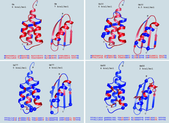
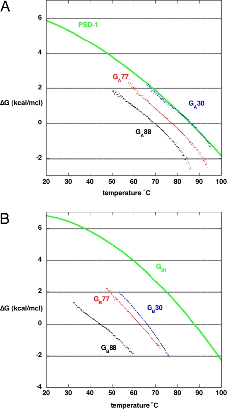
To identify a simplified code for conformational switching, we have redesigned two natural proteins to have 88% sequence identity but different tertiary structures: a 3-alpha helix fold and an alpha/beta fold. We describe the design of these homologous heteromorphic proteins, their structural properties as determined by NMR, their conformational stabilities, and their affinities for their respective ligands: IgG and serum albumin. Each of these proteins is completely folded at 25 degrees C, is monomeric, and retains the native binding activity. The complete binding epitope for both ligands is encoded within each of the proteins. The IgG-binding epitope is functional only in the alpha/beta fold, and the albumin-binding epitope is functional only in the 3-alpha fold. These results demonstrate that two monomeric folds and two different functions can be encoded with only 12% of the amino acids in a protein (7 of 56). The fact that 49 aa in these proteins are compatible with both folds shows that the essential information determining a fold can be highly concentrated in a few amino acids and that a very limited subset of interactions in the protein can tip the balance from one monomer fold to another. This delicate balance helps explain why protein structure prediction is so challenging. Furthermore, because a few mutations can result in both new conformation and new function, the evolution of new folds driven by natural selection for alternative functions may be much more probable than previously recognized.


Conflict of interest statement

The authors declare no conflict of interest.

Figures

Fig. 1.
Fig. 1.
Binding epitopes of GA and GB. (A) HSA-binding epitope of GA. Shown is a cartoon depiction of the secondary structure of GA. Amino acids making direct contacts with HSA (gray) are shown in yellow. Amino acids that were mutated to create a latent IgG epitope are in cyan (from Protein Data Bank entry 1TFO) (22). (B) IgG(Fc)-binding epitope of GB. Shown is a cartoon depiction of the secondary structure of GB. Amino acids making direct contacts with Fc (gray) are shown in cyan. Amino acids that were mutated to create a latent HSA epitope are in yellow (from Protein Data Bank entry 1fcc) (40).
Scheme. 1.
Scheme. 1.
Experimental design.
Scheme. 2.
Scheme. 2.
Experimental design.
Fig. 2.
Fig. 2.
Iterative design of heteromorphic pairs. For each heteromorphic pair, amino acid identities are shown in blue and nonidentities are shown in red. Mutations introduced in each design cycle are shown as blue spheres in the corresponding panel. The seven unique amino acids in GA88 and GB88 are shown as red sticks in Lower Right. Stabilities were measured in 0.1 M potassium phosphate buffer (pH 7.2) and extrapolated to 25°C (Fig. 4). The cartoons were generated from Protein Data Bank entries 2FS1 (PSD1) and 1PGA (GB1).
Fig. 3.
Fig. 3.
Analysis of conformation and thermal denaturation by CD. CD spectra and thermal denaturation curves are shown for GA30 (blue dashed line), GA77 (red dashed line), GA88 (black dashed line), GB30 (blue solid line), GB77 (red solid line), and GB88 (black solid line). (A) Mean residue ellipticity (degrees per cm2/dmol) is plotted vs. wavelength. Spectra were measured in 0.1 M potassium phosphate buffer (pH 7.2) using a 1-cm cylindrical cuvette at 25°C with [protein] = 5 μM. (B) Millidegrees at 222 nm are plotted vs. temperature in the range from 25°C to 100°C. The temperature profile was recorded by using a 1-cm cylindrical cuvette with a protein concentration of 5 μM in 0.1 M potassium phosphate buffer (pH 7.2).
Fig. 4.
Fig. 4.
Stability profiles of natural and mutant GA and GB proteins. (A) ΔG vs. temperature plots are shown for GA30 (blue), GA77 (red), and GA88 (black). For reference, the stability curve for the parent protein PSD1 is shown in green. (B) ΔG vs. temperature plots are shown for GB30 (blue), GB77 (red), and GB88 (black). For reference, the stability curve for the parent protein GB1 is shown in green.
Fig. 5.
Fig. 5.
1H, 15N HSQC spectra. Main chain amide assignments are shown for GA88 (black) and GB88 (red). Forty-nine of the 56 aa in these two proteins are identical but have different chemical environments reflecting the two different folds. Amide proton signals for side chains are connected by the horizontal lines. Chemical shift index data for GA88 and GB88 are included in SI Tables 1 and 2.

Similar articles

Cited by

References

    1. Fahnestock SR, Alexander P, Nagle J, Filpula D. J Bacteriol. 1986;167:870–880. - PMC - PubMed
    1. Falkenberg C, Bjork L, Åkerstrom B. 1992;31:1451–1457. - PubMed
    1. Frick IM, Wikstrom M, Forsen S, Drakenberg T, Gomi H, Sjobring U, Bjorck L. Proc Natl Acad Sci USA. 1992;89:8532–8536. - PMC - PubMed
    1. Myhre EB, Kronvall G. Infect Immun. 1977;17:475–482. - PMC - PubMed
    1. Reis KJ, Ayoub EM, Boyle MDP. J Immunol. 1984;132:3098–3102. - PubMed

Publication types

MeSH terms