. 2007 Jul 10;104(28):11748-53.
doi: 10.1073/pnas.0700019104. Epub 2007 Jul 3.

PIP5KI gamma is required for cardiovascular and neuronal development

Affiliations

PIP5KI gamma is required for cardiovascular and neuronal development

Yanfeng Wang et al. Proc Natl Acad Sci U S A. .

Abstract

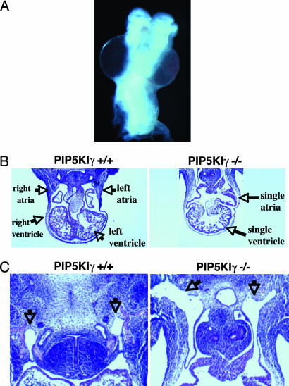
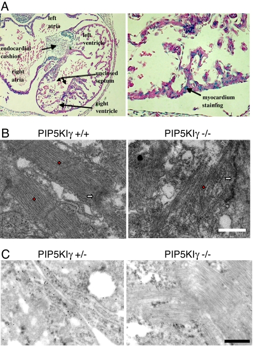
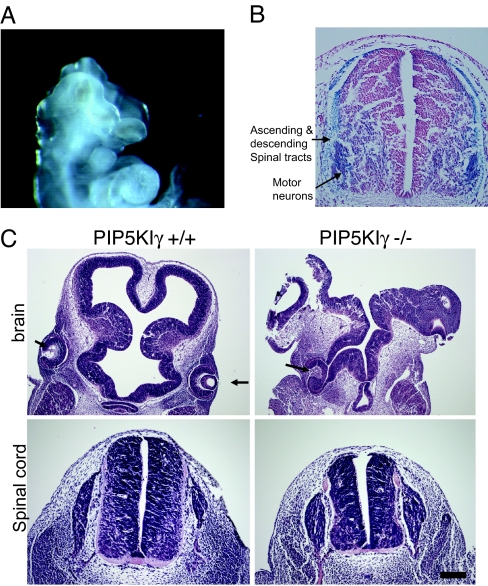
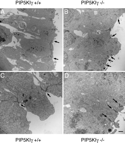
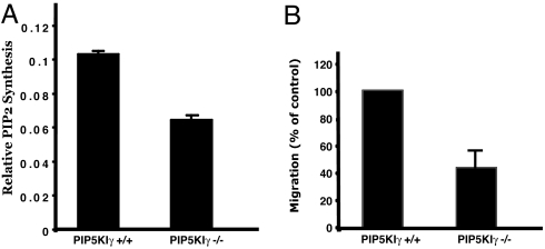
All eukaryotic cells contain the phospholipid phosphatidylinositol 4, 5-bisphosphate (PIP2) that serves multiple roles in signal transduction cascades. Type I phosphatidylinositol-4-phosphate 5-kinase (PIP5KI) catalyzes the synthesis of PIP2 by phosphorylating phosphatidylinositol 4 phosphate. Although the classical isoforms of PIP5KI (designated as alpha, beta, and gamma) all generate the same phospholipid product, they have significantly dissimilar primary structures and expression levels in different tissues, and they appear to localize within different compartments within the cell. Therefore, it appears likely that PIP5KI isoforms have overlapping, but not identical, functions. Here we show that targeted disruption of PIP5KIgamma causes widespread developmental and cellular defects. PIP5KIgamma-null embryos have myocardial developmental defects associated with impaired intracellular junctions that lead to heart failure and extensive prenatal lethality at embryonic day 11.5 of development. Loss of PIP5KIgamma also results in neural tube closure defects that were associated with impaired PIP2 production, adhesion junction formation, and neuronal cell migration. These data, along with those of other PIP5KI isoforms, indicate that individual PIP5KI isoenzymes fulfill specific roles in embryonic development.


Conflict of interest statement

The authors declare no conflict of interest.

Figures

Fig. 1.
Fig. 1.
Schematic of PIP5KIγ gene targeting. Diagram shows the location of the β-geo within the first intron of the PIP5KIγ gene. Location of the Southern blot probe is shown in blue. The gene traps insert an XbaI site. The insertion leads to a read-through mutation within the first intron of the targeted gene and truncates PIP5KIγ after the 32nd amino acid.
Fig. 2.
Fig. 2.
Loss of PIP5KIγ induces lethality at E11.5. (A) E9.5 PIP5KIγ−/− embryos are smaller compared to wild-type littermates (Left vs. Right). (B) The Southern blot of XbaI-digested DNA shows a 10.7-kb (wild-type) band and 4.5-kb (PIP5KIγ-targeted) band. Shown is an RT-PCR using a sense primer from exon 1 and antisense primers from β-geo and exon 3. Anti-PIP5KIγ immunoblot (BD Biosciences, San Jose, CA) shows complete loss of protein in brain lysates of knockout embryos.
Fig. 3.
Fig. 3.
PIP5KIγ-null embryos die of a cardiovascular defect. E11.5 PIP5KIγ-null embryos have a pericardial effusion (anterior view) (A), a single ventricle (B), and enlarged cardinal veins (C, arrows).
Fig. 4.
Fig. 4.
PIP5KIγ is required for actin organization and fascia adhesion formation in myocardiocytes. (A) E10.5 PIP5KIγ knockout embryo has an extremely atrophic right ventricle and a ventricular septum that has failed to close (Left). As seen by distribution of β-gal in the PIP5KIγ+/− heart shown, only myocardial cells are predicted to express PIP5KIγ mRNA (Right). (B) Electron micrographs of wild-type and PIP5KIγ-null cardiocytes. The red diamonds overlay the actin-rich sarcomeres, and the white arrows indicate the location of the fascia adherens. Loss of PIP5KIγ leads to actin disorganization, and the loss of the normal association of actin cables with the fascia adherens. (Scale bar: 1.0 μm.) (C) Immunogold-coupled anti-N-cadherin staining of PIP5KIγ+/− and PIP5KIγ−/− cardiocytes demonstrates that cells lacking PIP5KIγ do not have N-cadherin at the fascia adherens. (Scale bar: 500 nm.)
Fig. 5.
Fig. 5.
Neuronal defects in PIP5KIγ−/− embryos. (A) PIP5Kγ-null embryos fail to close their neuroepithelium. (B) Distribution of β-gal expression within the spinal cord of an E10.5 PIP5Kγ+/− embryo. This staining pattern predicts that PIP5Kγ is expressed most prominently in the motor column and the developing ascending and descending spinal tracts. (C) Cranial sections through telencephalon (Upper) and diencephalon (Lower) of E10.5 embryos at the level of the eyes (arrows.) The wild-type embryo displays normal development (Left). The PIP5KIγ−/− embryo (Right) has an unclosed neural tube and an abnormally organized neuroepithelium. Although the PIP5KIγ−/− cord has a normal general morphology, it is reduced in its dorsoventral axis and medial-lateral thickness. (Scale bar: 25 μm.)
Fig. 6.
Fig. 6.
Loss of PIP5Kγ impairs adherens junction formation in the brain. Electron micrographs from neuroepithelium of E8.5 PIP5KIγ+/+ (A and C) and PIP5KIγ−/− (B and D) embryos. Adherens junctions (arrows) are found between cells at the apical border. PIP5KIγ+/+ neuroepithelial cells exhibit longer and more complex branched junctions compared to PIP5KIγ−/− cells. (Scale bar: 2 μm.)
Fig. 7.
Fig. 7.
PIP5KIγ−/− neuronal cells exhibit defective PIP2 synthesis and cell migration. (A) Lysates of embryonic brains were analyzed for in vitro kinase activity by using PI4P as the exogenous substrate. (B) PIP5KIγ-null neuronal precursor cells have impaired migration in a transwell assay. Shown is mean ± SEM for three experiments (migration is normalized for control cells).

References

    1. Hokin MR, Hokin LE. J Biol Chem. 1953;203:967–977. - PubMed
    1. Toker A. Curr Opin Cell Biol. 1998;10:254–261. - PubMed
    1. Doughman RL, Firestone AJ, Anderson RA. J Membr Biol. 2003;194:77–89. - PubMed
    1. Ishihara H, Shibasaki Y, Kizuki N, Katagiri H, Yazaki Y, Asano T, Oka Y. J Biol Chem. 1996;271:23611–23614. - PubMed
    1. Loijens JC, Anderson RA. J Biol Chem. 1996;271:32937–32943. - PubMed

Publication types

MeSH terms

Substances