. 2007 Oct 1;110(7):2286-95.
doi: 10.1182/blood-2007-04-084996. Epub 2007 Jul 3.

Salinosporamide A (NPI-0052) potentiates apoptosis, suppresses osteoclastogenesis, and inhibits invasion through down-modulation of NF-kappaB regulated gene products

Affiliations

Salinosporamide A (NPI-0052) potentiates apoptosis, suppresses osteoclastogenesis, and inhibits invasion through down-modulation of NF-kappaB regulated gene products

Kwang Seok Ahn et al. Blood. .

Abstract

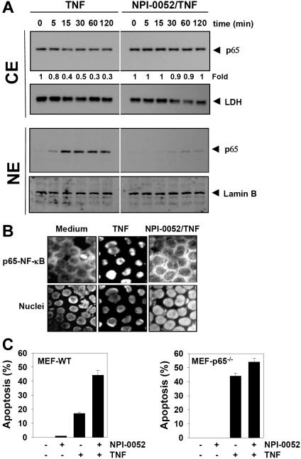
Salinosporamide A (also called NPI-0052), recently identified from the marine bacterium Salinispora tropica, is a potent inhibitor of 20S proteasome and exhibits therapeutic potential against a wide variety of tumors through a poorly understood mechanism. Here we demonstrate that salinosporamide A potentiated the apoptosis induced by tumor necrosis factor alpha (TNF), bortezomib, and thalidomide, and this correlated with down-regulation of gene products that mediate cell proliferation (cyclin D1, cyclooxygenase-2 [COX-2], and c-Myc), cell survival (Bcl-2, Bcl-xL, cFLIP, TRAF1, IAP1, IAP2, and survivin), invasion (matrix metallopro-teinase-9 [MMP-9] and ICAM-1), and angiogenesis (vascular endothelial growth factor [VEGF]). Salinosporamide A also suppressed TNF-induced tumor cell invasion and receptor activator of nuclear factor kappaB ligand (RANKL)-induced osteoclastogenesis. We also found that it suppressed both constitutive and inducible NF-kappaB activation. Compared with bortezomib, MG-132, N-acetyl-leucyl-leucyl-norleucinal (ALLN), and lactacystin, salinosporamide A was found to be the most potent suppressor of NF-kappaB activation. Further studies showed that salinosporamide A inhibited TNF-induced inhibitory subunit of NF-kappaB alpha (IkappaBalpha) degradation, nuclear translocation of p65, and NF-kappaB-dependent reporter gene expression but had no effect on IkappaBalpha kinase activation, IkappaBalpha phosphorylation, or IkappaBalpha ubiquitination. Thus, overall, our results indicate that salinosporamide A enhances apoptosis, suppresses osteoclastogenesis, and inhibits invasion through suppression of the NF-kappaB pathway.


Figures

Figure 1
Figure 1
NPI-0052 enhances apoptosis induced by TNF and chemotherapeutic drugs. (A) KBM-5 cells (10 000 cells/0.1 mL) were incubated at 37°C with 1 nM TNF, 10 μg/mL thalidomide, or 20 nM bortezomib in the presence and absence of 50 nM NPI-0052 as indicated for 18 hour, and the viable cells were assayed using the MTT reagent. The results are expressed as mean cytotoxicity (± standard deviation [SD]) from triplicate cultures. (B) Cells (106/mL) were pretreated with 50 nM NPI-0052 for 4 hours and then incubated with 1 nM TNF, 10 μg/mL thalidomide, or 20 nM bortezomib for 16 hours. Cell death was determined by the calcein-AM based live/dead assay as described in “Live and dead assay.” Data are representative of 3 independent experiments showing similar results. (C) U266 (106/mL) or DU145 (105/mL) cells were pretreated with 50 nM NPI-0052 for 4 hours and then incubated with 10 μg/mL thalidomide or 20 nM bortezomib for 16 hours. Cell death was determined by the calcein-AM based live/dead assay as described in “Live and dead assay.” Data are representative of 3 independent experiments showing similar results. Images were acquired as described in “Live and dead assay.” (D) Cells were pretreated with 50 nM NPI-0052 for 4 hours and then incubated with 1 nM TNF for 18 hours. Afterward, they were incubated with anti-annexin V antibody conjugated with FITC plus PI and analyzed with a flow cytometer for apoptotic effects. (E) Cells were pretreated with 50 nM NPI-0052 for 4 hours and then incubated with 1 nM TNF for 18 hours. Cells were fixed, stained with TUNEL assay reagent, and then analyzed with flow cytometer. (F) Cells were pretreated with 50 nM NPI-0052 for 4 hours and then incubated with 1 nM TNF for the indicated times. Whole-cell extracts were prepared and subjected to Western blot analysis using anti-PARP antibody.
Figure 2
Figure 2
NPI-0052 suppresses TNF-induced invasive activity and RANKL-induced osteoclastogenesis. (A) H1299 cells (2.5 × 10⁴) were seeded into the upper wells of a Matrigel invasion chamber overnight in the absence of serum, pretreated with 50 nM NPI-0052 for 4 hours, treated with 1 nM TNF for 24 hours in the presence of 5% serum, and then subjected to invasion assay. The value for no NPI-0052 and no TNF was set to 1.0. (B) RAW 264.7 cells (104) were plated overnight, pretreated with 50 nM NPI-0052 for 4 hours, and then treated with 5 nM RANKL. At 5 days later, cells were stained for TRAP and evaluated for osteoclastogenesis. Cells were analyzed under a phase contrast microscope (Nikon, Tokyo, Japan) and photographs were taken using Photometrics Coolsnap CF color camera (Nikon, Lewisville, TX). (C) Photographs were taken after 5 days of incubation with RANKL. The numbers of TRAP-positive multinucleated osteoclasts (> 3 nuclei) per well were counted. Error bars indicate SD of triplicate value.
Figure 3
Figure 3
NPI-0052 represses TNF-induced NF-κB–dependent expression of proliferation-, antiapoptosis-, and metastasis-related gene products. Proliferative (A), antiapoptotic (B), and metastatic (C) gene products are shown. KBM-5 cells were incubated with 50 nM NPI-0052 for 4 hours and then treated with 1 nM TNF for the indicated times. Whole-cell extracts were prepared, and 30 μg of the whole-cell lysate was resolved by SDS-PAGE, electrotransferred to nitrocellulose membrane, sliced from the membrane based on the molecular weight, and then probed with antibodies against survivin, IAP1/2, Bcl-2, Bcl-xL, cFLIP, TRAF1, VEGF, MMP-9, ICAM-1, COX-2, c-Myc, Cyclin D1, or β-actin as described in “Materials and methods.” TNF-treated and TNF plus NPI-0052-treated samples were run on the same gel under identical conditions and probed with the same immunoblotting solutions.
Figure 4
Figure 4
NPI-0052 inhibits both inducible and constitutive NF-κB activation in a dose- and time-dependent manner. (A) KBM-5 cells were incubated with the indicated concentrations of NPI-0052 for 4 hours and then exposed to 0.1 nM TNF for 30 minutes. The nuclear extracts were subjected to EMSA to evaluate NF-κB activation. (B) Cells were preincubated with 50 nM NPI-0052 for the indicated times, treated with 0.1 nM TNF for 30 minutes, and then subjected to EMSA to evaluate NF-κB activation. H1299 (C) or A293 (D) cells were pretreated with 50 nM NPI-0052 for 4 hours and then treated with 0.1 nM TNF for 30 minutes. The nuclear extracts were then prepared and assayed for NF-κB by EMSA as described in “Materials and methods.” (E) U266 cells with constitutive NF-κB activation cells were incubated with (±) 50 nM NPI-0052 for 12 hours. Nuclear extracts were prepared and analyzed for NF-κB activation by EMSA.
Figure 5
Figure 5
NPI-0052 inhibits TNF-dependent NF-κB activation and IκBα degradation. (A) Cells were preincubated with 50 nM NPI-0052 for 4 hours, treated with 0.1 nM TNF for the indicated times, and then subjected to EMSA and Western blot analysis to evaluate NF-κB activation. (B) Cells were incubated with 50 nM NPI-0052 for 4 hours and then treated with 0.1 nM TNF for the indicated times. Cytoplasmic extracts were prepared, fractionated on SDS-PAGE, and electrotransferred to a nitrocellulose membrane. Western blot analysis was performed with anti-IκBα and anti-phospho-specific anti-IκBα. (C) Cells were preincubated with 50 nM NPI-0052 for 4 hours, incubated with 50 μg/mL N-acetyl-leucyl-leucyl-norleucinal (ALLN) for 30 minutes, and then treated with 0.1 nM TNF for 15 minutes. Cytoplasmic extracts were fractionated and then subjected to Western blot analysis using phospho-specific anti-IκBα antibody. The same membrane was reblotted with anti-IκBα antibody. (D) Cells were preincubated with 50 nM NPI-0052 for 4 hours and then treated with 0.1 nM TNF for 15 minutes. Cytoplasmic extracts were immunoprecipitated with antibody against IκBα then subjected to Western blot analysis using monoclonal anti-ubiquitin antibody. (E) Cells were preincubated with 50 nM NPI-0052 for 4 hours and then treated with 1 nM TNF for the indicated times. Whole-cell extracts (1 mg/mL) were immunoprecipitated with antibody against IKK-α and analyzed using an immunocomplex kinase assay. Data are for a representative experiment of 3 independent ones showing similar results.
Figure 6
Figure 6
NPI-0052 inhibits TNF-induced p65 nuclear translocation. (A) KBM-5 cells were incubated with 50 nM NPI-0052 for 4 hours and then treated with 0.1 nM TNF for the indicated times. Cytoplasmic and nuclear extracts were prepared, fractionated on SDS-PAGE, and electrotransferred to a nitrocellulose membrane. The analysis was performed using p65 antibodies. (B) Immunocytochemical analysis of TNF-induced p65 nuclear translocation. KBM-5 cells were incubated with 50 nM NPI-0052 for 4 hours, treated with 1 nM TNF for 15 minutes, and then subjected to immunocytochemical analysis as described in “Immunolocalization of NF-κB p65.” Cells plated with Mounting Media (Sigma-Aldrich) were analyzed under a fluorescence microscope (Lapshot-2, Nikon) equipped with a CFWN 10 /1.5 NA oil-immersion objective lens and a Photometrics Coolsnap CF color camera (Nikon). Images were acquired with MetaMorph 4.6.5 software (Universal Imaging). (C) The wild-type and p65−/− (105/mL) cells were pretreated with 50 nM NPI-0052 for 4 hours and then incubated with 1 nM TNF for 16 hours. Cell death was determined by the calcein-AM based live/dead assay as described in “Live and dead assay.” Data are for a representative experiment of 3 independent ones showing similar results. Error bars represent SD of triplicate values.
Figure 7
Figure 7
NPI-0052 inhibits TNF-induced NF-κB–dependent reporter gene (SEAP) expression and suppresses chymotrypsin-like activity of 20S proteasome. (A) A293 cells were transiently transfected with an NF-κB–containing plasmid linked to the SEAP gene and then pretreated with the indicated concentrations of NPI-0052 for 4 hours. After 24 hours in culture with 1 nM TNF, cell supernatants were collected and assayed for SEAP activity as described in “Materials and methods.” Results are expressed as fold activity over the activity of the vector control. (B) Chemical structures of various proteasome inhibitors. (C) KBM-5 (1 × 10⁶) cells were incubated with 50 nM each of various proteasome inhibitors for 4 hours and then exposed to 0.1 nM TNF for 30 minutes. The nuclear extracts were subjected to EMSA to evaluate NF-κB activation. (D) RPMI 8226 and PC-3 cells were treated with various concentrations of NPI-0052 and bortezomib for 1 hour, prepared the cell lysates, and assayed for the chymotrypsin-like activity of the 20S proteasome as described in “Materials and methods.” Results are presented as percentage inhibition compared with the chymotrypsin-like activity observed in untreated cells. Error bars represent SD of triplicate values.

References

    1. Newman DJ, Cragg GM, Snader KM. Natural products as sources of new drugs over the period 1981-2002. J Nat Prod. 2003;66:1022–1037. - PubMed
    1. Feling RH, Buchanan GO, Mincer TJ, Kauffman CA, Jensen PR, Fenical W. Salinosporamide A: a highly cytotoxic proteasome inhibitor from a novel microbial source, a marine bacterium of the new genus Salinospora. Angew Chem Int Ed Engl. 2003;42:355–357. - PubMed
    1. Jensen PR, Mincer TJ, Williams PG, Fenical W. Marine actinomycete diversity and natural product discovery. Antonie Van Leeuwenhoek. 2005;87:43–48. - PubMed
    1. Macherla VR, Mitchell SS, Manam RR, et al. Structure-activity relationship studies of Salinosporamide A (NPI-0052), a novel marine derived proteasome inhibitor. J Med Chem. 2005;48:3684–3687. - PubMed
    1. Groll M, Huber R, Potts BC. Crystal structures of Salinosporamide A (NPI-0052) and B (NPI-0047) in complex with the 20S proteasome reveal important consequences of beta-lactone ring opening and a mechanism for irreversible binding. J Am Chem Soc. 2006;128:5136–5141. - PubMed

Publication types

MeSH terms