

Systemic distribution of West Nile virus infection: postmortem immunohistochemical study of six cases
- PMID: 17610522
- PMCID: PMC8095553
- DOI: 10.1111/j.1750-3639.2007.00080.x
Systemic distribution of West Nile virus infection: postmortem immunohistochemical study of six cases
Abstract
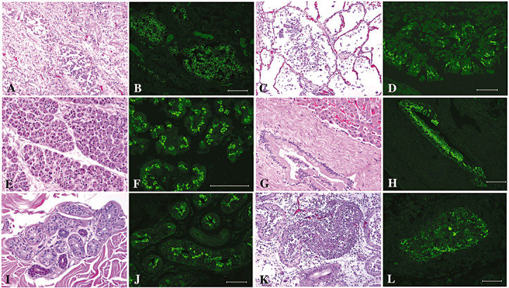
Rare cases of West Nile virus (WNV)-associated inflammation outside the central nervous system (CNS) have been reported. We evaluated the systemic distribution of WNV in postmortem tissues during encephalitis in six patients using immunohistochemistry. WNV antigens were detected in neurons of CNS (all 6 cases), kidney (4 cases), lungs (2 cases), pancreas (2 cases), thyroid (2 cases), intestine (2 cases), stomach (1 case), esophagus (1 case), bile duct (1 case), skin (1 case), prostate (1 case) and testis (1 case). In systemic organs epithelial cells were infected. In none of the six cases were viral antigens identified in hepatocytes, heart, adrenal gland, nerves, skeletal muscles, bone, vessels and fat. All cases in which viral antigens were identified in systemic organs in addition to CNS were severely immunocompromised transplant recipients. With the exception of testis and brain, most foci of infection were not associated with inflammation. While the absence of inflammation may in part be due to patient immunosuppression or to possible transient nature of any host response, compartmentalization of viral antigen to the luminal region of epithelial cells may sequester WNV from immune recognition. Comparison of our findings with previous reports suggests that patients with WNV encephalitis can have widespread systemic infection.
Figures


References
-
- Agamanolis DP, Leslie MJ, Caveny EA, Guarner J, Shieh WJ, Zaki SR (2003) Neuropathological findings in West Nile virus encephalitis: a case report. Ann Neurol 54:547–551. - PubMed
-
- Anninger WV, Lomeo MD, Dingle J, Epstein AD, Lubow M (2003) West Nile virus‐associated optic neuritis and chorioretinitis. Am J Ophthalmol 136:1183–1185. - PubMed
-
- Armstrong WS, Bashour CA, Smedira NG, Heupler FA, Hoeltge GA, Mawhorter SD, Sudheendra V, Gordon SM (2003) A case of fatal West Nile virus meningoencephalitis associated with receipt of blood transfusions after open heart surgery. Ann Thorac Surg 76:605–607. - PubMed
-
- Barshes NR, Agee EE, Zgabay T, Brunicardi FC, Goss JA, Debakey ME (2006) West Nile virus encephalopathy following pancreatic islet transplantation. Am J Transplant 6:3037. - PubMed
Publication types
MeSH terms
Substances
Grants and funding
LinkOut - more resources
Full Text Sources
Medical
