

Functional MRI of working memory and selective attention in vibrotactile frequency discrimination
- PMID: 17610721
- PMCID: PMC1925104
- DOI: 10.1186/1471-2202-8-48
Functional MRI of working memory and selective attention in vibrotactile frequency discrimination
Abstract
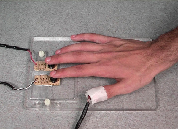
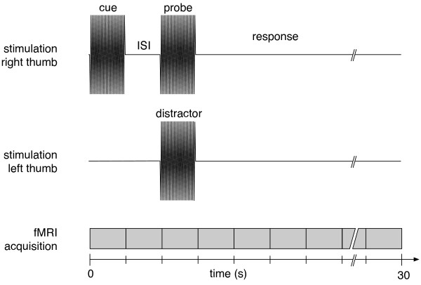
Background: Focal lesions of the frontal, parietal and temporal lobe may interfere with tactile working memory and attention. To characterise the neural correlates of intact vibrotactile working memory and attention, functional MRI was conducted in 12 healthy young adults. Participants performed a forced-choice vibrotactile frequency discrimination task, comparing a cue stimulus of fixed frequency to their right thumb with a probe stimulus of identical or higher frequency. To investigate working memory, the time interval between the 2 stimuli was pseudo-randomized (either 2 or 8 s). To investigate selective attention, a distractor stimulus was occasionally presented contralaterally, simultaneous to the probe.
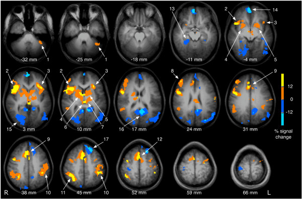
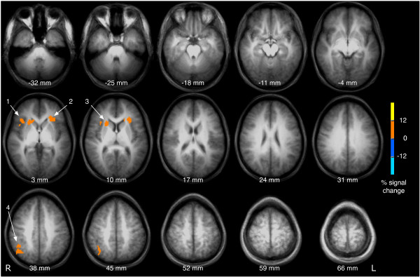
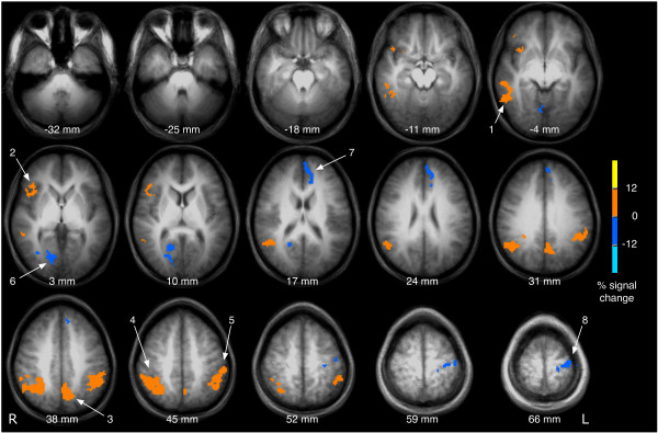
Results: Delayed vibrotactile frequency discrimination, following a probe presented 8 s after the cue in contrast to a probe presented 2 s after the cue, was associated with activation in the bilateral anterior insula and the right inferior parietal cortex. Frequency discrimination under distraction was correlated with activation in the right anterior insula, in the bilateral posterior parietal cortex, and in the right middle temporal gyrus.
Conclusion: These results support the notion that working memory and attention are organised in partly overlapping neural circuits. In contrast to previous reports in the visual or auditory domain, this study emphasises the involvement of the anterior insula in vibrotactile working memory and selective attention.
Figures
Similar articles
-
Neural correlates of spatial working memory in humans: a functional magnetic resonance imaging study comparing visual and tactile processes.Neuroscience. 2006 Apr 28;139(1):339-49. doi: 10.1016/j.neuroscience.2005.08.045. Epub 2005 Dec 1. Neuroscience. 2006. PMID: 16324793
-
Sequential neural processes of tactile-visual crossmodal working memory.Neuroscience. 2006 Apr 28;139(1):299-309. doi: 10.1016/j.neuroscience.2005.05.058. Epub 2005 Dec 1. Neuroscience. 2006. PMID: 16324794
-
Oscillatory correlates of vibrotactile frequency processing in human working memory.J Neurosci. 2010 Mar 24;30(12):4496-502. doi: 10.1523/JNEUROSCI.6041-09.2010. J Neurosci. 2010. PMID: 20335486 Free PMC article.
-
Working memory for order information: multiple cognitive and neural mechanisms.Neuroscience. 2006 Apr 28;139(1):195-200. doi: 10.1016/j.neuroscience.2005.08.024. Epub 2005 Dec 15. Neuroscience. 2006. PMID: 16359810 Review.
-
Posterior parietal cortex: an interface between attention and learning?Neurobiol Learn Mem. 2009 Feb;91(2):114-20. doi: 10.1016/j.nlm.2008.07.004. Epub 2008 Aug 15. Neurobiol Learn Mem. 2009. PMID: 18675370 Free PMC article. Review.
Cited by
-
Interoception and drug addiction.Neuropharmacology. 2014 Jan;76 Pt B(0 0):342-50. doi: 10.1016/j.neuropharm.2013.07.002. Epub 2013 Jul 12. Neuropharmacology. 2014. PMID: 23855999 Free PMC article. Review.
-
Indoor apparent temperature, cognition, and daytime sleepiness among low-income adults in a temperate climate.Indoor Air. 2022 Jan;32(1):e12972. doi: 10.1111/ina.12972. Epub 2021 Dec 9. Indoor Air. 2022. PMID: 34888941 Free PMC article.
-
Levodopa and the feedback process on set-shifting in Parkinson's disease.Hum Brain Mapp. 2012 Jan;33(1):27-39. doi: 10.1002/hbm.21187. Epub 2011 Mar 24. Hum Brain Mapp. 2012. PMID: 21438075 Free PMC article.
-
Prefrontal and posterior parietal contributions to the perceptual awareness of touch.Sci Rep. 2019 Nov 18;9(1):16981. doi: 10.1038/s41598-019-53637-w. Sci Rep. 2019. PMID: 31740713 Free PMC article.
-
Irrelevant sensory stimuli interfere with working memory storage: evidence from a computational model of prefrontal neurons.Cogn Affect Behav Neurosci. 2013 Mar;13(1):23-34. doi: 10.3758/s13415-012-0131-9. Cogn Affect Behav Neurosci. 2013. PMID: 23138530
References
-
- Gardner E, Kandel E. Touch. In: Kandel E, Schwartz J, Jessell T, editor. Principles of neural science. New York: McGraw-Hill; 2000. pp. 451–471.
-
- Chao LL, Knight RT. Human prefrontal lesions increase distractibility to irrelevant sensory inputs. Neuroreport. 1995;6:1605–1610. - PubMed
Publication types
MeSH terms
Grants and funding
LinkOut - more resources
Full Text Sources
Other Literature Sources
Medical
