. 2007 Jul 4:8:49.
doi: 10.1186/1471-2202-8-49.

TRPM5, a taste-signaling transient receptor potential ion-channel, is a ubiquitous signaling component in chemosensory cells

Affiliations

TRPM5, a taste-signaling transient receptor potential ion-channel, is a ubiquitous signaling component in chemosensory cells

Silke Kaske et al. BMC Neurosci. .

Abstract

Background: A growing number of TRP channels have been identified as key players in the sensation of smell, temperature, mechanical forces and taste. TRPM5 is known to be abundantly expressed in taste receptor cells where it participates in sweet, amino acid and bitter perception. A role of TRPM5 in other sensory systems, however, has not been studied so far.

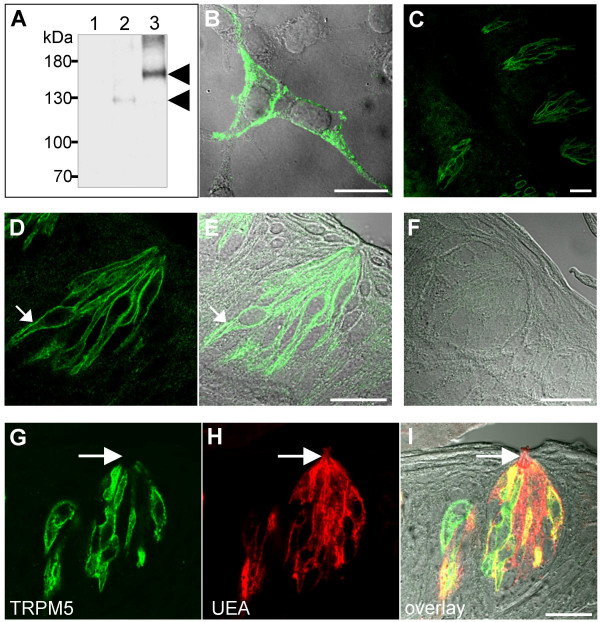
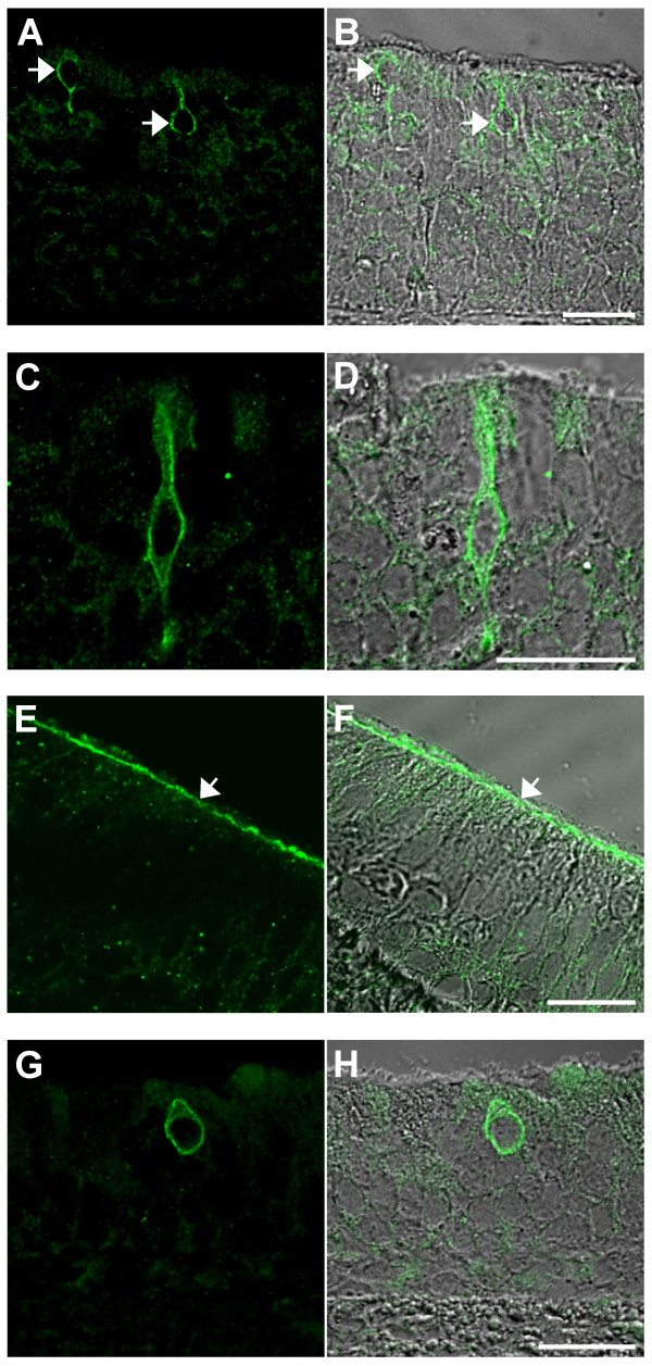
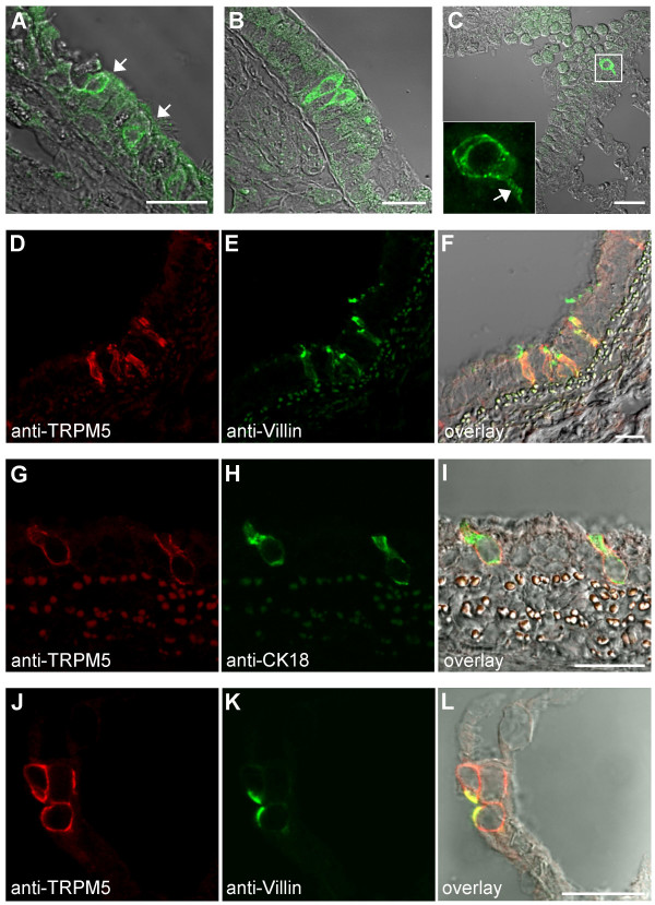
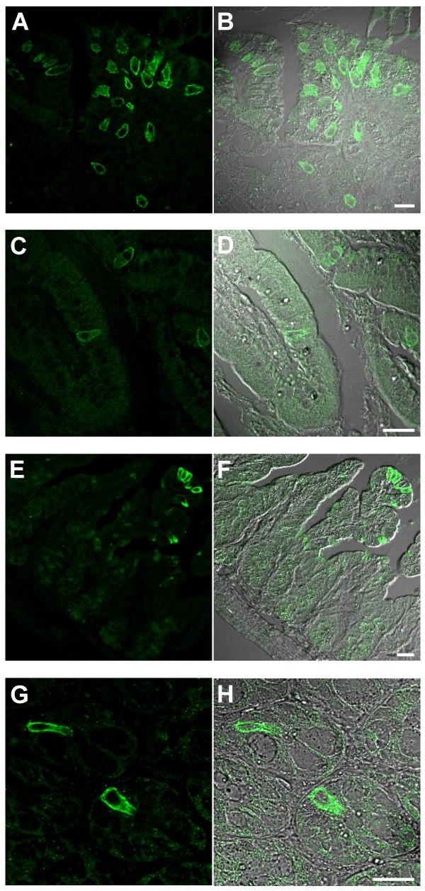
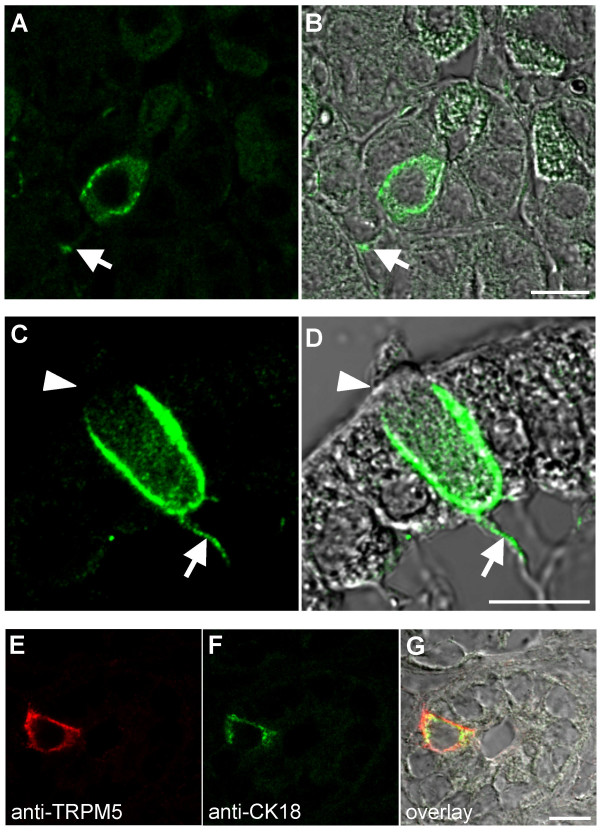
Results: Here, we systematically investigated the expression of TRPM5 in rat and mouse tissues. Apart from taste buds, where we found TRPM5 to be predominantly localized on the basolateral surface of taste receptor cells, TRPM5 immunoreactivity was seen in other chemosensory organs - the main olfactory epithelium and the vomeronasal organ. Most strikingly, we found solitary TRPM5-enriched epithelial cells in all parts of the respiratory and gastrointestinal tract. Based on their tissue distribution, the low cell density, morphological features and co-immunostaining with different epithelial markers, we identified these cells as brush cells (also known as tuft, fibrillovesicular, multivesicular or caveolated cells). In terms of morphological characteristics, brush cells resemble taste receptor cells, while their origin and biological role are still under intensive debate.

Conclusion: We consider TRPM5 to be an intrinsic signaling component of mammalian chemosensory organs, and provide evidence for brush cells being an important cellular correlate in the periphery.


Figures

Figure 1
Figure 1
Immunolocalization of TRPM5 in taste buds of mouse papillae vallatae. The TRPM5-specific antibody Ab-321 recognizes TRPM5 transiently expressed in HEK 293 cells. (A) Western blot analysis of total lysates of mock- (line 1), TRPM5- (line 2) and YFP-TRPM5- (line 3) transfected HEK 293 cells. Positions of TRPM5- and YFP-TRPM5-specific bands are indicated by arrowheads. (B) Overlay of confocal and corresponding differential interference contrast (DIC) images of immunofluorescence staining of HEK 293 cells transiently expressing wild-type TRPM5. (C-E) Immunohistochemistry of taste buds using a TRPM5-specific antibody at lower (C) and higher magnifications (D, E). Notably, TRPM5 is predominantly localized on the basolateral surface of cells (indicated by the arrow in (D, E)). (F) Lack of TRPM5 immunoreactivity in taste receptors after preincubation of the TRPM5-specific antibody with the immunization peptide. (G-I) Fluorescent labeling of taste buds with the TRPM5-specific antibody (E) and the UEA lectin (H) and overlay of both with corresponding DIC image (I). Note that only a subset of cells in the taste bud is enriched in TRPM5. In the TRPM5-positive cells, TRPM5 is mainly localized on the basolateral cell surface and absent in microvilli (as indicated by an arrow (G, H, I)). Scale bars are 20 μm each.
Figure 2
Figure 2
TRPM5 expression in mouse main olfactory epithelium and the vomeronasal organ. Confocal images of TRPM5-specific signals (left panels) and overlay with corresponding DIC images (right panel) are depicted. (A-D) Immunolocalization of TRPM5 in solitary cells of the main olfactory epithelium is shown at lower (indicated by the arrows in (A, B)) and higher magnifications (C, D). (E-H) Immunohistochemistry of TRPM5 in the VNO. (E, F) Localization of TRPM5 on the apical surface of sensory epithelia (indicated by the arrow). (G, H) Detection of TRPM5-enriched solitary cells in the non-sensory part of the VNO. Scale bars are 20 μm.
Figure 3
Figure 3
TRPM5 is specifically expressed in brush cells of the respiratory system. Detection of solitary TRPM5-expressing cells in the mouse respiratory epithelium of nose (indicated by arrows in (A)), trachea (B) and bronchus (C). Overlays of TRPM5 fluorescence with the corresponding DIC images are shown. The inset in (C) shows the fluorescent image of a TRPM5-positive cell with a basolateral extension (indicated by the arrow). (D-L) TRPM5-expressing cells express brush cell-specific marker proteins. Confocal images of TRPM5-specific signals (left panels), villin or CK18 (middle panels) and their overlays with corresponding DIC images (right panel) are shown. Note the co-localization of TRPM5 with villin (D-F) and CK18 (G-H) in rat trachea and TRPM5 with villin in rat alveolar epithelium (J-L). Scale bars are 20 μm.
Figure 4
Figure 4
Distribution of TRPM5-immunoreactive cells in the mouse gastrointestinal tract. Confocal images of TRPM5-specific signals (left panel) and their overlay with the corresponding DIC images (right panel) are depicted. (A, B) TRPM5-expressing cells are abundant in the cardiac region of the stomach. In gut epithelia, TRPM5-enriched cells are detected in the duodenal villi (C, D), the ileum (E, F) and the colon (G, H). Scale bars are 20 μm.
Figure 5
Figure 5
TRPM5-expressing cells of the gastrointestinal tract have morphological and biochemical characteristics of brush cells. (A-D) Morphological features of TRPM5-expressing cells. Confocal images of TRPM5-specific signals (left panels) and overlay with corresponding DIC images (right panels) are shown. The TRPM5-enriched cells in gastric glands of mouse stomach (A, B) and in mouse a duodenal villus (C, D) reveal a long basolateral processes (indicated by the arrows). Within duodenal epithelia (C, D), the TRPM5-immunoreactive cell contains a protrusion extending beyond the epithelial luminal border (indicated by an arrowhead). (E-G) Co-localization of TRPM5 with CK18 in intestinal glands of the rat duodenum. Confocal images of TRPM5 and CK18 specific signals (E, F) and their overlay with corresponding DIC images (G) are depicted. Scale bars are 10 μm.

Similar articles

Cited by

References

    1. Dhaka A, Viswanath V, Patapoutian A. TRP Ion Channels and Temperature Sensation. Annu Rev Neurosci. 2006;29:135–161. doi: 10.1146/annurev.neuro.29.051605.112958. - DOI - PubMed
    1. Ramsey IS, Delling M, Clapham DE. An introduction to TRP channels. Annu Rev Physiol. 2006;68:619–647. doi: 10.1146/annurev.physiol.68.040204.100431. - DOI - PubMed
    1. Liman ER, Corey DP, Dulac C. TRP2: a candidate transduction channel for mammalian pheromone sensory signaling. Proc Natl Acad Sci U S A. 1999;96:5791–5796. doi: 10.1073/pnas.96.10.5791. - DOI - PMC - PubMed
    1. Stowers L, Holy TE, Meister M, Dulac C, Koentges G. Loss of sex discrimination and male-male aggression in mice deficient for TRP2. Science. 2002;295:1493–1500. doi: 10.1126/science.1069259. - DOI - PubMed
    1. Huang AL, Chen X, Hoon MA, Chandrashekar J, Guo W, Trankner D, Ryba NJ, Zuker CS. The cells and logic for mammalian sour taste detection. Nature. 2006;442:934–938. doi: 10.1038/nature05084. - DOI - PMC - PubMed

Publication types

MeSH terms

LinkOut - more resources