Comparative Study
. 2007 Jul 4;8(1):47.
doi: 10.1186/1465-9921-8-47.

Phenotypic alterations in type II alveolar epithelial cells in CD4+ T cell mediated lung inflammation

Affiliations
Comparative Study

Phenotypic alterations in type II alveolar epithelial cells in CD4+ T cell mediated lung inflammation

Marcus Gereke et al. Respir Res. .

Abstract

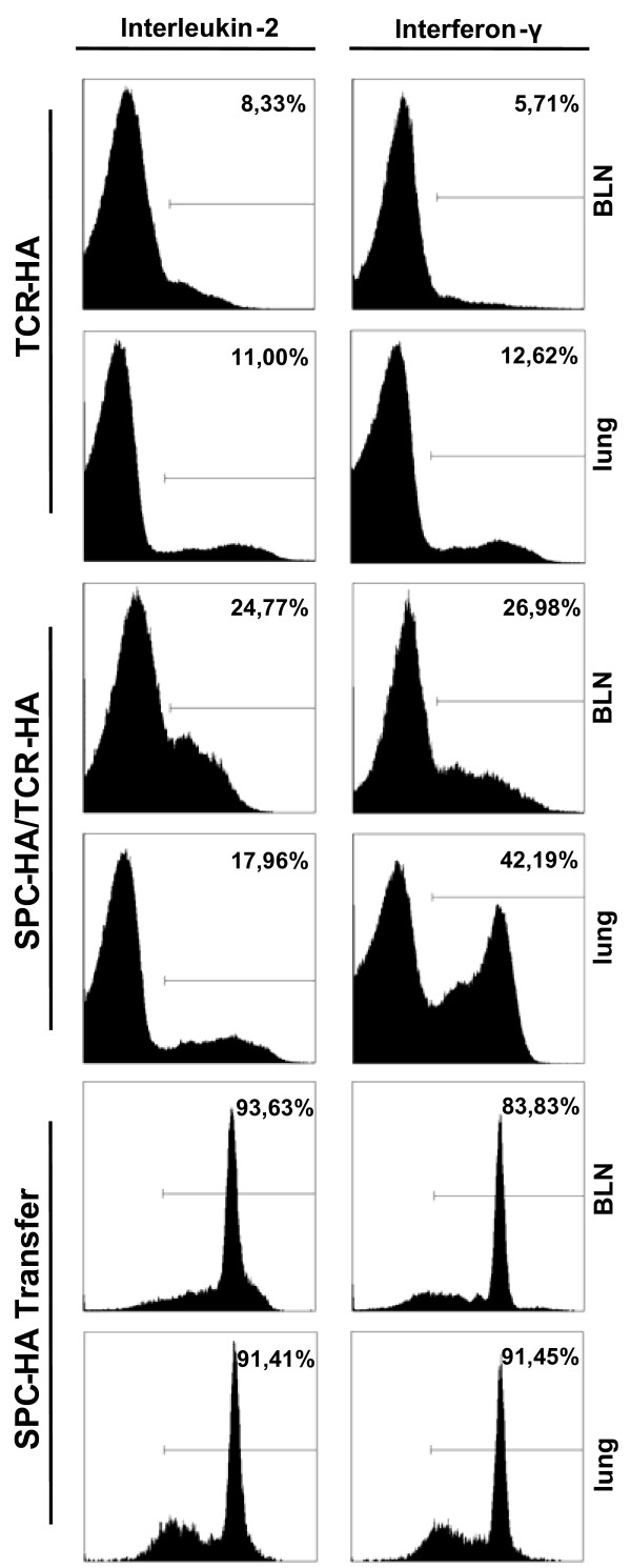
Background: Although the contribution of alveolar type II epithelial cell (AEC II) activities in various aspects of respiratory immune regulation has become increasingly appreciated, our understanding of the contribution of AEC II transcriptosome in immunopathologic lung injury remains poorly understood. We have previously established a mouse model for chronic T cell-mediated pulmonary inflammation in which influenza hemagglutinin (HA) is expressed as a transgene in AEC II, in mice expressing a transgenic T cell receptor specific for a class II-restricted epitope of HA. Pulmonary inflammation in these mice occurs as a result of CD4+ T cell recognition of alveolar antigen. This model was utilized to assess the profile of inflammatory mediators expressed by alveolar epithelial target cells triggered by antigen-specific recognition in CD4+ T cell-mediated lung inflammation.

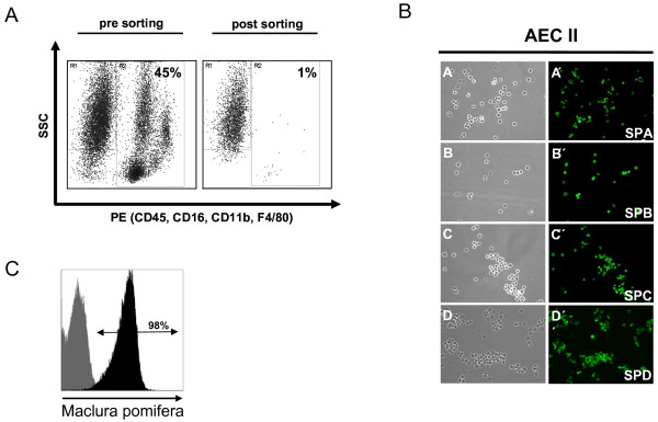
Methods: We established a method that allows the flow cytometric negative selection and isolation of primary AEC II of high viability and purity. Genome wide transcriptional profiling was performed on mRNA isolated from AEC II isolated from healthy mice and from mice with acute and chronic CD4+ T cell-mediated pulmonary inflammation.

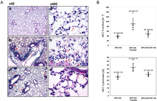
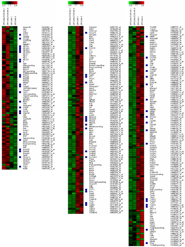
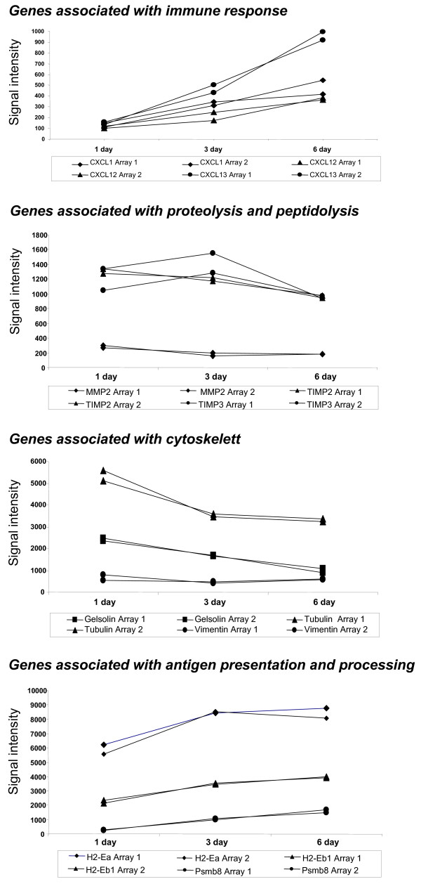
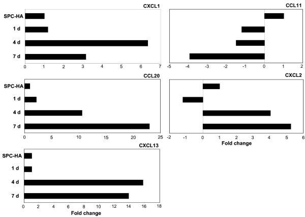
Results: T cell-mediated inflammation was associated with expression of a broad array of cytokine and chemokine genes by AEC II cell, indicating a potential contribution of epithelial-derived chemoattractants to the inflammatory cell parenchymal infiltration. Morphologically, there was an increase in the size of activated epithelial cells, and on the molecular level, comparative transcriptome analyses of AEC II from inflamed versus normal lungs provide a detailed characterization of the specific inflammatory genes expressed in AEC II induced in the context of CD4+ T cell-mediated pneumonitis.

Conclusion: An important contribution of AEC II gene expression to the orchestration and regulation of interstitial pneumonitis is suggested by the panoply of inflammatory genes expressed by this cell population, and this may provide insight into the molecular pathogenesis of pulmonary inflammatory states. CD4+ T cell recognition of antigen presented by AEC II cells appears to be a potent trigger for activation of the alveolar cell inflammatory transcriptosome.


Figures

Figure 1
Figure 1
CD4+ T cell recognition of alveolar epithelial antigen results in airway inflammation and AEC II hypertrophy. (A) Histological examination of lungs from healthy SPC-HA (a and a'), SPC-HA six days after adoptive transfer of HA-specific CD4+ T cells (b, b') and SPC-HA/TCR-HA double transgenic mice (c, c'). Lung sections were stained with H&E. Black arrows indicate AEC II, red arrows indicate lymphocytes. No lesions were detectable in the lung of SPC-HA mice. Specifically, type II pneumocytes were completely unchanged (a, a'). A moderate, perivascular and peribronchiolar infiltration with mature lymphocytes was detected in the lung of SPC-HA mice after transfer with HA-specific CD4+ T cells. Adjacent to these infiltrations, a slight connective tissue edema and a mild infiltration with neutrophils were observed. Type II pneumocytes in the vicinity of the lymphocytic infiltrations were moderately hypertrophic. A few alveolar macrophages were present in the alveoli (b, b'). Moderate, multifocal, perivascular and peribronchiolar infiltrations with lymphocytes were present in the lung of SPC-HA/TCR-HA double transgenic mice. Type II pneumocytes close to the lymphocytic infiltrations were mildly activated and hypertrophic (c, c'). (B) Histological results were corroborated morphometrically by measuring AEC II surface and perimeter to quantify the degree of cellular hypertrophy (n = 15, 3 mice with 5 AEC II per mouse; ± standard deviation). AEC II surface: SPC-HA vs SPC-HA Transfer: P < 0,001), SPC-HA vs SPC-HA/TCR-HA (P < 0,0001), SPC-HA transfer vs SPC-HA/TCR-HA (P < 0,0001). AEC II perimeter: SPC-HA vs SPC-HA Transfer: P < 0,001), SPC-HA vs SPC-HA/TCR-HA (P < 0,001), SPC-HA transfer vs SPC-HA/TCR-HA (P < 0,001). All Student's t-test.
Figure 2
Figure 2
Intracellular cytokine staining in CD4+ T cells. CD4+ T cells from the lung or bronchial lymph nodes (BLN) from either TCR-HA control mice, SPC-HA/TCR-HA double transgenic mice or SPC-HA mice adoptively transferred with HA-specific CD4+ T cells were analyzed by FACS for the expression of interleukin 2 and interferon γ.
Figure 3
Figure 3
Purification of alveolar type II epithelial cells by fluorescence-activated cell sorting. (A) Cell suspension obtained by enzymatic tissue disintegration and subsequent sequential filtration was labelled with antibodies to CD45, CD16, CD32, CD11b, and F4/80. Antibody negative AEC II were further distinguished from other cells by size and granularity. Reanalysis of sorted cells demonstrated an extremely low frequency of contaminating hematopoetic cells. (B) Sorted cells express surfactant proteins A, B, C and D. Cytospins of sorted AEC II cells were stained for the surfactant proteins A, B, C and D. Almost all cells were found to be positive for all four surfactant proteins. A, B, C and D represent phase contrast microscopy, A', B', C', and D' represent immunohistochemical stainings for the corresponding surfactant protein. (C) Staining of sorted AEC II with Maclura pomifera lectin revealed high purity of isolated cells. Black histogram indicates staining with the lectin, grey histogram indicates unstained cells.
Figure 4
Figure 4
Heat map including genes differentially expressed in AEC II cells isolated from lungs of diseased SPC-HA/TCR-HA as well as healthy SPC-HA mice. Red indicates induction of gene expression, green indicates repression (+2: bright red; -2: bright green). Black indicates no changes. Blue squares indicate genes further highlighted in Table 1. Genes were considered to be regulated whose expression was at least twofold increased or decreased.
Figure 5
Figure 5
Time course of gene expression in AEC II after adoptive CD4+ T cell transfer into SPC-HA mice. AEC II cells were isolated from the lung of SPC-HA mice one (n = 3), three (n = 3) and six (n = 3) days after adoptive transfer of HA-specific CD4+ T cells. Cells were subjected to microarray analysis and the level of gene expression over time is depicted for selected genes. Data obtained from two different experiments are represented.
Figure 6
Figure 6
Chemokine expression in AEC II after adoptive CD4+ T cell transfer into SPC-HA mice. AEC II cells were isolated from the lung of SPC-HA mice one (n = 3), three (n = 3) and six (n = 3) days after adoptive transfer of HA-specific CD4+ T cells. Cells were subjected to quantitative real-time RT-PCR analyses. mRNA expression levels of CXCL1, CCL20, CXCL13, CCL11, CXCL2, and RPS9 (as internal control) were analyzed in real-time RT-PCR assays. Relative mRNA amounts were normalized with respect to expression levels in AEC II cells isolated from SPC-HA mice not receiving CD4+ T cell transfer (fold change = 1).

References

    1. Folkerts G, Nijkamp FP. Airway epithelium: more than just a barrier! Trends Pharmacol Sci. 1998;19:334–341. doi: 10.1016/S0165-6147(98)01232-2. - DOI - PubMed
    1. Knight DA, Holgate ST. The airway epithelium: structural and functional properties in health and disease. Respirology. 2003;8:432–446. doi: 10.1046/j.1440-1843.2003.00493.x. - DOI - PubMed
    1. Crapo JD, Barry BE, Gehr P, Bachofen M, Weibel ER. Cell number and cell characteristics of the normal human lung. Am Rev Respir Dis. 1982;126:332–337. - PubMed
    1. Kalina M, Mason RJ, Shannon JM. Surfactant protein C is expressed in alveolar type II cells but not in Clara cells of rat lung. Am J Respir Cell Mol Biol. 1992;6:594–600. - PubMed
    1. Lesur O, Arsalane K, Lane D. Lung alveolar epithelial cell migration in vitro: modulators and regulation processes. Am J Physiol. 1996;270:L311–L319. - PubMed

Publication types