. 2007 Oct 25;367(2):307-23.
doi: 10.1016/j.virol.2007.05.041. Epub 2007 Jul 5.

Alpha-beta T cells provide protection against lethal encephalitis in the murine model of VEEV infection

Affiliations

Alpha-beta T cells provide protection against lethal encephalitis in the murine model of VEEV infection

Slobodan Paessler et al. Virology. .

Abstract

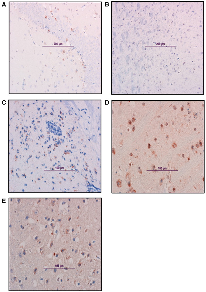
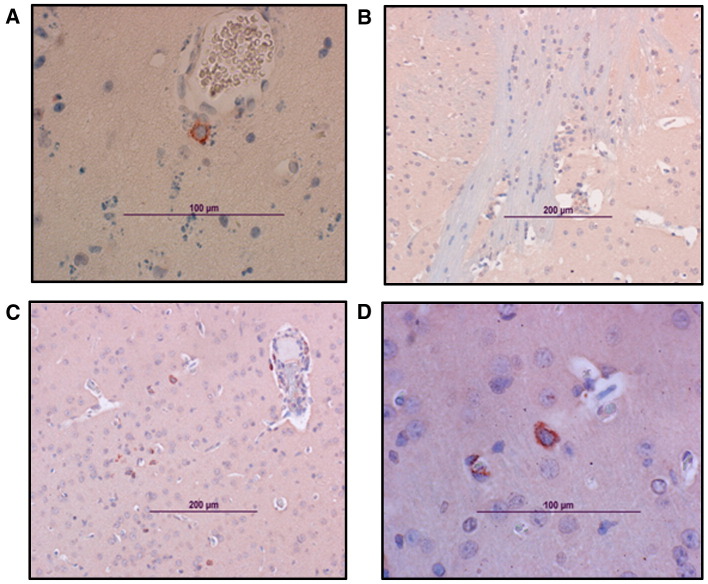
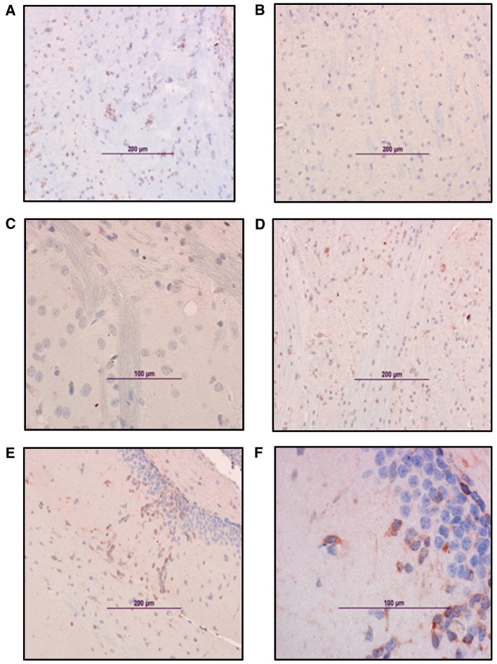
We evaluated the safety and immunogenicity of a chimeric alphavirus vaccine candidate in mice with selective immunodeficiencies. This vaccine candidate was highly attenuated in mice with deficiencies in the B and T cell compartments, as well as in mice with deficient gamma-interferon responsiveness. However, the level of protection varied among the strains tested. Wild type mice were protected against lethal VEEV challenge. In contrast, alpha/beta (alphabeta) TCR-deficient mice developed lethal encephalitis following VEEV challenge, while mice deficient in gamma/delta (gammadelta) T cells were protected. Surprisingly, the vaccine potency was diminished by 50% in animals lacking interferon-gamma receptor alpha chain (R1)-chain and a minority of vaccinated immunoglobulin heavy chain-deficient (microMT) mice survived challenge, which suggests that neutralizing antibody may not be absolutely required for protection. Prolonged replication of encephalitic VEEV in the brain of pre-immunized mice is not lethal and adoptive transfer experiments indicate that CD3(+) T cells are required for protection.


Figures

Fig. 1
Fig. 1
Vaccine safety studies in mice. Survival and average body temperature of wild type and immunodeficient mice vaccinated with live-attenuated chimeric alphavirus vaccine (SIN/ZPC). Panels A and B show the survival and average body temperature of parental C57BL/6 (wild type, WT) and immunodeficient mice inoculated with live-attenuated chimeric alphavirus vaccine (SIN/ZPC). Panel C shows the average body temperature following VEEV infection of unvaccinated mice. The total percentage of surviving mice (A) and the average body temperature (B) of SIN/ZPC vaccinated IFNγR KO, γδ TCR KO, μMT KO and WT mice before and after vaccination on day 0 were evaluated, as follows. Vaccination: eleven to 18-week-old, female mice were inoculated on day 0 and day 34 subcutaneously into the medial thigh with the chimeric SIN/ZPC virus at a dose of 5 × 105 plaque forming units in a total volume of 100 μl. Mock-vaccinated mice (indicated as “WT* unvacc”) were inoculated subcutaneously with 100 μl of PBS. Virus challenge: SIN/ZPC or mock-vaccinated mice were inoculated on day 0 intranasally with an identical dose of virulent VEEV (ZPC738) in 40 μl of PBS and deaths were recorded. Daily telemetric monitoring of body temperature was performed for a total period of 14 days, beginning 2 days prior to the first vaccination on day 0. Asterisk () indicates parental C57BL/6 mice (wild type, WT). The characteristics of the parental WT and immunodeficient mice are described in Materials and methods (Table 1). (C) Positive control for telemetric monitoring. The body temperature of unvaccinated γδ TCR KO mice 2 days before (day − 1 and − 2) and 7 days after (day + 1 through + 7) infection with virulent VEEV (ZPC738) on day 0.
Fig. 2
Fig. 2
Vaccine efficacy in the mouse model. Survival of SIN/ZPC double-vaccinated wild type and immunodeficient mice following challenge with virulent VEEV. Six-week-old, female parental C57BL/6 (wild type, WT) and immunodeficient mice were vaccinated on day − 42 and day − 14 subcutaneously into the medial thigh with the chimeric SIN/ZPC vaccine at a dose of 5 × 105 plaque forming units (PFU) in a total volume of 100 μl of PBS or with PBS alone (WT unvacc). Deaths were recorded following intranasal challenge with virulent VEEV (ZPC738) at a dose of 2 × 105 PFU per animal (ca. 1000 LD50) in 20 μl of PBS at 6 weeks following initial vaccination. Asterisk () indicates parental C57BL/6 mice (wild type, WT) and the characteristics of immunodeficient mice are described in Materials and methods (Table 1). (A) The total percentage of surviving mice. (B) Number of surviving mice in replicate experiments. Comparison of survival curves using the logrank test (Altman, 1991) indicates that there is a statistically significant difference in survival overall between the experimental groups over the 28-day monitoring period (logrank, p < 0.0001). Fisher's exact test was used to compare the survival proportion of each vaccinated strain to the mock vaccinated WT, and the individual p-values are indicated on the survival curve.
Fig. 3
Fig. 3
Vaccine efficacy in the mouse model. Mean VEEV titer in organs of SIN/ZPC vaccinated wild type and immunodeficient mice at 3 and 7 days post-challenge with ZPC738. Mice were immunized on day − 42 and day − 14 with chimeric SIN/ZPC vaccine at a dose of 5 × 105 plaque-forming units (PFU) in 100 μl of PBS or with PBS (WT* Mock) via subcutaneous route and challenged intranasally with 2 × 105 PFU of virulent VEEV (ZPC738) per animal in 20 μl of PBS. Asterisk () indicates parental C57BL/6 mice (wild type, WT) and the characteristics of immunodeficient mice are described in Materials and methods (Table 1). Animals were sacrificed at (A) 3 days and (B) 7 days post-challenge and organs were harvested for viral titration. The limit of detection was 40 PFU/g, as indicated by the solid line.
Fig. 4
Fig. 4
Vaccine efficacy in the mouse model. Histopathological analysis of brains from vaccinated mice challenged intranasally with VEEV ZPC738. Brains were harvested from mice obtained following the vaccination and challenge experiments described in Figs. 2 and3. The characteristics of parental C57BL/6 mice (wild type, WT) and immunodeficient mice are described in Materials and methods (Table 1). Photomicrographs of hematoxylin and eosin-stained brains obtained on day 7 from the following mice: (A) WT unvaccinated, (B) WT vaccinated; and the following chimeric SIN/ZPC-vaccinated immunodeficient mice: (C) μMT, (D) αβ TCR KO, and (E) γδ TCR KO mice. (F) A higher magnification image of panel E is shown. Multifocal mononuclear cell infiltrates as well as focal necrosis and neuronal cell death characterized through hyperangulation of hypereosinophilic neurons and loss of neuropil, are prominent in γδ TCR KO and αβ TCR KO mice. μMT KO mice have prominent micro-hemorrhages in addition to inflammation.
Fig. 5
Fig. 5
Vaccine efficacy in the mouse model. Immunohistochemical analysis of the brain from SIN/ZPC- or mock-vaccinated and VEEV-challenged mice. Viral antigen in the brain. Representative photomicrographs were obtained from immunohistological analysis of brains of 6-day-old female chimeric SIN/ZPC virus- or mock-vaccinated mice euthanized on day 7 following virulent VEEV (ZPC738) challenge infection, as described in Figs. 2 and3. The characteristics of parental C57BL/6 mice (wild type, WT) and immunodeficient mice are described in Materials and methods (Table 1). The localization of viral antigen (reddish-brown) was demonstrated via immunostaining of brain sections using antibody to VEEV (ATCC, cat number: VR1250AF). VEEV antibody was detected using biotinylated secondary antibody, followed by avidin–peroxidase color development. Slides were counter-stained with Mayer's Modified Hematoxylin prior to mounting and microscopy. (A) WT unvaccinated, (B) WT chimeric SIN/ZPC-vaccinated; and the following chimeric SIN/ZPC-vaccinated immunodeficient mice: (C) μMT, (D) αβ TCR KO mice and (E) γδ TCR KO. Viral antigen is detected in association with neuronal cells in the brain stem (C and D), hippocampus (A) and cortex (E).
Fig. 6
Fig. 6
Vaccine efficacy in the mouse model. Immunohistochemical analysis of the brain from SIN/ZPC- and mock-vaccinated and VEEV-challenged mice. B cells in the brain. Brains were harvested from mice obtained following the vaccination and challenge experiments described in Figs. 2 and3. The characteristics of parental C57BL/6 mice (wild type, WT) and immunodeficient mice are described in Materials and methods (Table 1). B cells were detected by staining with antibody to the B cell marker, CD45R/B220 and counter-stained with Mayer's Modified Hematoxylin prior to mounting and microscopy, as described in Materials and methods. CD45R/B220-positive cells (reddish-brown) in brains of chimeric SIN/ZPC-vaccinated, virulent VEEV (ZPC738) challenged animals from the following immunodeficient mice are shown: (A) αβ TCR KO, 7 days post-infection (dpi), (B) γδ TCR KO mice, 7 dpi, (C) γδ TCR KO, 12 dpi, and (D) a higher magnification image of panel C.
Fig. 7
Fig. 7
Vaccine efficacy in the mouse model. Immunohistochemical analysis of the brain from SIN/ZPC- and mock-vaccinated and VEEV-challenged mice. T cells in the brain. Brains were harvested from mice obtained following the vaccination and challenge experiments described in Figs. 2 and3. The characteristics of parental C57BL/6 mice (wild type, WT) and immunodeficient mice are described in Materials and methods (Table 1). T cells were detected by staining with antibody to the T cell marker, CD3, and counter-stained with Mayer's Modified Hematoxylin prior to mounting and microscopy, as described in Materials and methods. CD3-positive cells (reddish-brown) are shown in the brains of the following animals: (A) WT mock-vaccinated; 7 days post-infection (dpi), (B) WT chimeric SIN/ZPC-vaccinated; 7 dpi, and for the following vaccinated immunodeficient mice: (C) αβ TCR KO at 7 dpi, (D) γδ TCR KO at 7 dpi, (E) γδ TCR KO at 12 dpi. (F) A higher magnification image of panel E is shown.
Fig. 8
Fig. 8
Vaccine efficacy in a single immunization model and protection mediated by passive cell transfer. (A) Six-week-old, female γδ TCR-deficient mice were vaccinated on day − 42 into the medial thigh with the chimeric SIN/ZPC vaccine at a dose of 5 × 105 plaque forming units (PFU) in a total volume of 100 μl of PBS or with PBS alone (WT mock). Deaths were recorded following intranasal challenge with virulent VEEV (ZPC738) at a dose of 2 × 105 PFU per animal (ca. 1000 LD50) in 20 μl of PBS at 6 weeks following initial vaccination. Asterisk () indicates parental C57BL/6 mice (wild type, WT) and the characteristics of immunodeficient mice are described in Materials and methods (Table 1). (B–C) Naïve (recipient) αβ TCR-deficient mice (N = 15) were injected on day − 28 via intraperitoneal route with 5 × 107 CD3+ T cells (isolated as described in Materials and methods) suspended in 100 μl of sterile phosphate-buffered saline (PBS). As controls for reconstitution experiments, five mice were inoculated with PBS alone. Recipient mice were immunized with chimeric SIN/ZPC 2 days after T cell transfer and challenged with virulent VEEV (ZPC738, 2 × 105 PFU/animal) at 28 days following immunization. Animals were monitored for death, as indicated. Due to Hurricane Rita evacuation, experiment shown in 2A was prematurely terminated at 6 days post-challenge.

Similar articles

Cited by

References

    1. Aaronson R.P., Young J.F., Palese P. Oligonucleotide mapping: evaluation of its sensitivity by computer-simulation. Nucleic Acids Res. 1982;10:237–246. - PMC - PubMed
    1. Agrati C., Castilletti C., De Santis R., Cimini E., Bordi L., Malkovsky M., Poccia F., Capobianchi M.R. Interferon- gamma -mediated antiviral immunity against orthopoxvirus infection is provided by gamma delta T cells. J. Infect. Dis. 2006;193(11):1606–1607. - PubMed
    1. Altman D.G. Practical Statistics for Medical Research. Chapman and Hall; London: 1991. Analysis of survival times.
    1. Anishchenko M., Bowen R.A., Paessler S., Austgen L., Greene I.P., Weaver S.C. Venezuelan encephalitis emergence mediated by a phylogenetically predicted viral mutation. Proc. Natl. Acad. Sci. U.S.A. 2006;103(13):4994–4999. - PMC - PubMed
    1. Beaty B.J., Calisher C.H., Shope R.E. Arboviruses. In: Schmidt N.J., Emmons R.W., editors. Diagnostic Procedures for Viral, Rickettsial and Chlamydial Infections. 6th edition. American Public Health Association; Washington, DC: 1989. pp. 797–855.

Publication types

MeSH terms

Substances