Comparative Study
. 2007 Jul 4;27(27):7318-28.
doi: 10.1523/JNEUROSCI.1831-07.2007.

Phosphorylation of huntingtin by cyclin-dependent kinase 5 is induced by DNA damage and regulates wild-type and mutant huntingtin toxicity in neurons

Affiliations
Comparative Study

Phosphorylation of huntingtin by cyclin-dependent kinase 5 is induced by DNA damage and regulates wild-type and mutant huntingtin toxicity in neurons

Sandrine L Anne et al. J Neurosci. .

Abstract

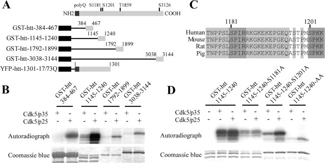
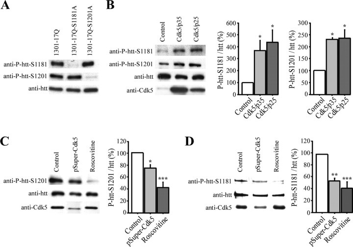
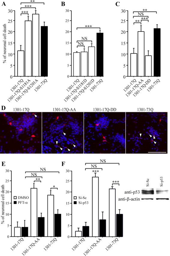
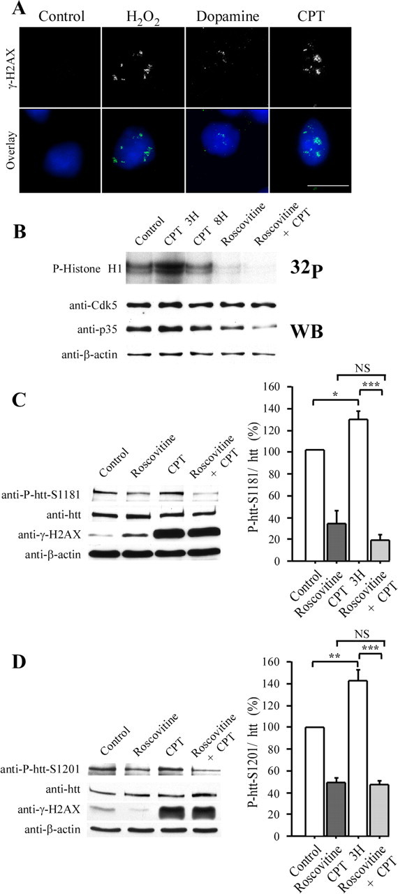
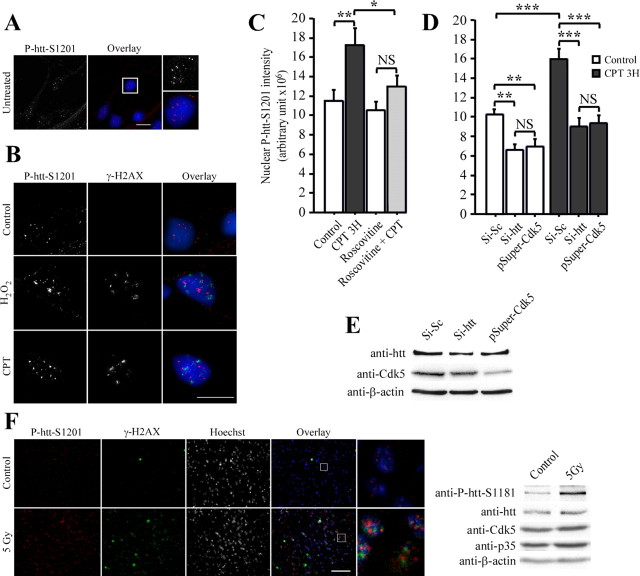
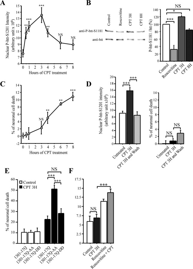
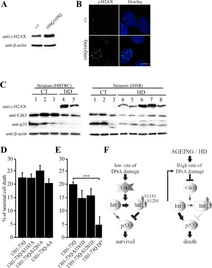
Huntingtin is an antiapoptotic protein that becomes toxic when its polyglutamine stretch is expanded, resulting in Huntington's disease (HD). Protein context and posttranslational modifications regulate huntingtin toxicity. Identifying signaling pathways that act on huntingtin is, therefore, key to understanding huntingtin function in normal and pathological conditions. We show here that huntingtin is phosphorylated by the cyclin-dependent kinase 5 (Cdk5) at serines 1181 and 1201. Phosphorylation can be induced by DNA damage in vitro and in vivo. The state of huntingtin phosphorylation is a crucial regulator of neuronal cell death. Absence of phosphorylation of huntingtin at serines 1181 and 1201 confers toxic properties to wild-type huntingtin in a p53-dependent manner in striatal neurons and accelerates neuronal death induced by DNA damage. In contrast, phosphorylation at serines 1181 and 1201 protects against polyQ-induced toxicity. Finally, we show in late stages of HD a sustained DNA damage that is associated with a decrease in Cdk5/p35 levels. We propose that wild-type huntingtin is a component of the DNA damage response signal in neurons and that the Cdk5/DNA damage pathway is dysregulated in HD.


Figures

Figure 1.
Figure 1.
Cdk5 phosphorylates huntingtin at S1181 and S1201 in vitro. A, The human htt sequence contains four consensus sites for Cdk5 phosphorylation. GST-fused htt and huntingtin 1301 fragments used in this study are shown. B, Several fragments of htt are phosphorylated by Cdk5 in vitro. Kinase assays are performed using recombinant Cdk5/p35 and Cdk5/p25 and GST-fused proteins as substrates. C, The Cdk5 consensus site is defined by S/T-P-X-K/R with phosphorylation on the S/T. It is well conserved in vertebrates for serines 1181 and 1201 of htt sequence. D, Serines 1181 and 1201 are phosphorylated in vitro. GST–htt–1145–1240 fragments with S1181A and S1201A mutations are subjected to phosphorylation by recombinant Cdk5/p35 and Cdk5/p25. YFP, Yellow fluorescent protein.
Figure 2.
Figure 2.
Cdk5 phosphorylates huntingtin at S1181 and S1201 in neurons. A–D, Immunoblotting experiments are performed using the anti-P-htt–S1181, anti-P-htt–S1201, anti-htt (4C8), anti-Cdk5, anti-p35, and anti-β-actin antibodies as indicated. A, To assess the specificity of the phospho-antibodies, 1301–17Q, 1301–17Q–S1181A, and 1301–17Q–S1201A are expressed in HEK 293T cells. B, Cdk5/p35 and Cdk5/p25 are expressed in HEK 293T cells. Quantification reveals a statistically significant increase in phosphorylation at S1181 and S1201 of endogenous htt in the presence of Cdk5/p35 and Cdk5/p25. C, D, Htt phosphorylation at S1181 and S1201 is significantly inhibited by decreasing Cdk5 levels by RNA interference (pSuper–Cdk5) or by roscovitine treatment. SH-SY5Y cells (C) and primary cultures of striatal neurons (D) were transfected with pSuper–Cdk5 or treated with roscovitine (10 μm, 3 h). Quantifications reveal decreased phosphorylations when Cdk5 activity is diminished. *p < 0.05; **p < 0.01; ***p < 0.001.
Figure 3.
Figure 3.
Absence of phosphorylation at serines 1181 and 1201 turns huntingtin into a toxic protein through a p53-dependent mechanism. A–F, Primary cultures of striatal neurons are transfected by a modified phosphate calcium technique with constructs encoding the first 1301 amino acids of human htt containing 17 (wild-type, 1301–17Q) or 73 (1301–73Q) glutamines with intact serines or serines to alanines or aspartic acids mutations. Neurons are scored for neuronal cell death. D, Striatal neurons are immunostained using anti-htt 4C8 antibody (red). Nuclei are stained with Hoechst. White arrows indicate degenerating neurons. Scale bar, 100 μm. E, F, p53 mediates polyQ-induced toxicity and the toxicity of wild-type htt induced by the absence of phosphorylations at serines 1181 and 1201. The activity of p53 is inhibited using pifithrin-α (PTF-α) (E) or RNA interference (F). Although a control siRNA (Si-Sc) had no effect, the p53-directed siRNA (Si-p53) lowers p53 levels as revealed by immunoblotting against p53 and β-actin (loading control). *p < 0.05; **p < 0.01; ***p < 0.001. NS, Not significant.
Figure 4.
Figure 4.
Neuronal DNA damage increases Cdk5 activity and phosphorylations of huntingtin at serines 1181 and 1201. A, Treatment of primary cultures of striatal neurons with DMSO (0.1%, control), 50 μm H2O2, 500 μm dopamine, and 10 μm CPT leads to DNA damage. Cells are immunostained for γ-H2AX. The nuclear staining is shown in blue. Scale bar, 10 μm. B, Cdk5 activity is elevated after 3 h of CPT treatment, although its activity returned to basal level after longer treatment. In vitro kinase assays are performed using immunoprecipitated Cdk5 from primary cultures of striatal neurons treated with DMSO (0.1%, control), 10 μm CPT (3 and 8 h), and, when indicated, 10 μm roscovitine (4 h). Histone H1 is used as a substrate. Analysis of the total extracts by anti-Cdk5, anti-p35, and anti-β-actin immunoblotting reveals that the increase in Cdk5 activity is not linked to an increase in Cdk5 and p35 levels. C, D, Primary cultures of striatal neurons (C) and SH-SY5Y cells (D) are treated with DMSO (0.1%, control), roscovitine (10 μm, 4 h), and 10 μm CPT (3 h). Protein extracts are analyzed by immunoblotting with anti-P-htt–S1181, anti-P-htt–S1201, anti-htt, anti-γ-H2AX, and anti-β-actin antibodies. C, D, Quantifications reveal significantly increased phosphorylations at S1181–S1201 during DNA damage and decreased phosphorylations when Cdk5 activity is diminished by roscovitine treatment. CPT is not able to induce phosphorylation of htt when cells are treated with roscovitine. *p < 0.05; **p < 0.01; ***p < 0.001. WB, Western blot; NS, not significant.
Figure 5.
Figure 5.
Phosphorylation of huntingtin at serine 1201 is induced by Cdk5 during DNA damage in cells and in vivo. A, B, P-htt–S1201-immunopositive nuclear staining is increased by DNA damage. Striatal neurons are untreated or treated with DMSO (0.1%, control), H2O2 (50 μm, 15 min) and CPT (10 μm, 3 h). Fixed neurons are immunostained for the presence of P-htt–S1201 and γ-H2AX. Nuclei are stained with Hoechst. Scale bars, 10 μm. C, D, P-htt–S1201-immunopositive nuclear staining is specific to Cdk5 and htt. Neurons are treated with DMSO (0.1%, control), roscovitine (10 μm, 4 hr), CPT (10 μm, 3 hr), and/or electroporated with control siRNA (Si-Sc), siRNAs directed against htt (Si-htt), or pSuper–Cdk5. Striatal neurons are fixed in methanol and immunostained for P-htt–S1201, and quantification of the nuclear P-htt–S1201 dots intensity is performed. E, Immunoblotting analysis revealed that si-htt and pSuper–Cdk5 reduce htt and Cdk5 levels, respectively, in primary cultures of striatal neurons. F, Radiation induces DNA damage and phosphorylation of htt at serines 1181 and 1201 in mouse. Striatal coronal sections of a wild-type mice 8 h after irradiation (bottom row) and of an unirradiated control (top row) were stained for the presence of P-htt–S1201 and γ-H2AX. Nuclei are stained with Hoechst. Scale bar, 100 μm. Brain extracts of an unirradiated control (Control) and of a wild-type mice 8 h after irradiation (5 Gy) were analyzed by immunoblotting for P-htt–S1181, htt, Cdk5, p35, and β-actin. *p < 0.05; **p < 0.01; ***p < 0.001. NS, Not significant.
Figure 6.
Figure 6.
Phosphorylation of huntingtin occurs early before neuronal death. A, Striatal neurons treated with CPT (10 μm) for varying lengths of time are fixed in methanol and immunostained for P-htt–S1201. Quantification of the nuclear P-htt–S1201 dots intensity reveals an increased phosphorylation at S1201 of htt at 3 h of CPT treatment. At 8 h, S1201 phosphorylation returns to basal level. B, Phosphorylation of htt at S1181 follows the same kinetic during CPT treatment than that of S1201. Primary culture of striatal neurons were treated with DMSO (0.1%, control), roscovitine (10 μm, 4 h), and CPT (10 μm, 3 and 8 h). Protein extracts of neuronal cultures were analyzed by immunoblotting with anti-P-S1181 and anti-htt antibodies. C, Neuronal cell death is induced at 8 h of CPT treatment. Neurons are treated as in A and fixed and scored for neuronal cell death. D, Neurons are treated with CPT (10 μm) for 3 h and washed with culture media for 5 h when indicated (wash), fixed in methanol, and immunostained for P-htt–S1201 or scored for neuronal death. The increased phosphorylation at S1201 of htt at 3 h of CPT treatment is reversible by removal of CPT. As a control, cell death remains constant. E, Unphosphorylated htt accelerates DNA damage-induced cell death. Neurons electroporated with low amount of 1301–17Q, 1301–17Q–AA, and 1301–17Q–DD and treated with DMSO (0.1%, control) and CPT (10 μm, 3 h) were scored for neuronal cell death. F, Reducing Cdk5 activity increases CPT-induced neuronal death. Striatal neurons were treated with DMSO (0.1%, control), CPT (10 μm, 3 h), and roscovitine (10 μm, 4 h) and analyzed as in A. *p < 0.05; **p < 0.01; ***p < 0.001. NS, Not significant.
Figure 7.
Figure 7.
DNA damage, cdk5, and phosphorylations of huntingtin at S1181 and S1201 in HD. A, B, Extracts from wild-type (+/+) and 109Q/109Q mouse striatal cells are analyzed for the presence of γ-H2AX. Immunoblotting with an anti-β-actin antibody is used as a control. Accumulation of γ-H2AX is observed in polyQ cells. B, Immunostaining of γ-H2AX nuclear foci in +/+ and 109Q/109Q cells reveals an increase of foci in the presence of polyQ–htt. Scale bar, 10 μm. C, Whereas γ-H2AX levels are increased in the striatum of HD patients, Cdk5 and p35 levels are reduced. Protein extracts are prepared from whole striatum of control (CT) and HD individuals and analyzed for the presence of γ-H2AX, Cdk5, and p35. Immunoblotting with an anti-β-actin antibody is used as a control. D, E, Phosphorylation of serines 1181 and 1201 in polyQ–htt blocks neuronal death. Primary cultures of striatal neurons are transfected by a modified phosphate calcium method with constructs encoding the first 1301 amino acids of human htt containing 73 glutamines (polyQ, 1301–73Q) with intact serines or serines to alanines or aspartic acids mutations. Neurons are scored for neuronal cell death. F, In normal conditions, DNA damage events occur from spontaneous decay, replication errors, and cellular metabolism. Cdk5 is then activated as a response to DNA damage leading to phosphorylation of serines 1181 and 1201 of htt. Htt phosphorylation blocks polyQ-induced neuronal death in a p53-dependent manner. In contrast, in aging and late stages of HD that correspond to accumulation of DNA damage, the activity of Cdk5 is decreased, and the reduced phosphorylation of wild-type htt turns it into a toxic protein, therefore accelerating DNA damage-induced cell death. ***p < 0.001.

References

    1. Bae BI, Xu H, Igarashi S, Fujimuro M, Agrawal N, Taya Y, Hayward SD, Moran TH, Montell C, Ross CA, Snyder SH, Sawa A. p53 mediates cellular dysfunction and behavioral abnormalities in Huntington's disease. Neuron. 2005;47:29–41. - PubMed
    1. Benchoua A, Trioulier Y, Zala D, Gaillard MC, Lefort N, Dufour N, Saudou F, Elalouf JM, Hirsch E, Hantraye P, Deglon N, Brouillet E. Involvement of mitochondrial complex II defects in neuronal death produced by N-terminus fragment of mutated huntingtin. Mol Biol Cell. 2006;17:1652–1663. - PMC - PubMed
    1. Bezprozvanny I, Hayden MR. Deranged neuronal calcium signaling and Huntington disease. Biochem Biophys Res Commun. 2004;322:1310–1317. - PubMed
    1. Bogdanov MB, Andreassen OA, Dedeoglu A, Ferrante RJ, Beal MF. Increased oxidative damage to DNA in a transgenic mouse model of Huntington's disease. J Neurochem. 2001;79:1246–1249. - PubMed
    1. Borrell-Pages M, Zala D, Humbert S, Saudou F. Huntington's disease: from huntingtin function and dysfunction to therapeutic strategies. Cell Mol Life Sci. 2006;63:2642–2660. - PMC - PubMed

Publication types

MeSH terms