Characterization of neurons in the CA2 subfield of the adult rat hippocampus
- PMID: 17611285
- PMCID: PMC6794598
- DOI: 10.1523/JNEUROSCI.1829-07.2007
Characterization of neurons in the CA2 subfield of the adult rat hippocampus
Abstract
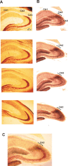
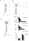
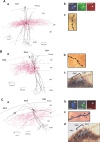
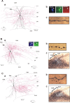
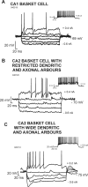
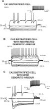
The hippocampal cornu ammonis 2 (CA2) region is unique in being the only CA region receiving inputs from the hypothalamic supramammillary nucleus, of importance in modulating hippocampal theta rhythm, and is seizure resistant in temporal lobe epilepsy. CA2 has, however, been little studied, possibly because of its small size and difficulty encountered in defining its borders. To investigate the properties of CA2 interneurons, intracellular recordings with biocytin filling were made in adult hippocampal slices. Two types of basket cells were identified. A minority resembled those in CA1, with fast spiking behavior, vertically oriented dendrites, and axons confined to the region of origin. In contrast, the majority of parvalbumin-immunopositive CA2 basket and bistratified cells had long, horizontally oriented, sparsely spiny dendrites extending into all CA subfields in stratum oriens, adapting firing patterns and a pronounced "sag" in voltage responses to hyperpolarizing current, indicative of I(h). Broad CA2 basket cells innervated all three CA subfields and could thus provide CA1 and CA2 with feedforward and CA3 with feedback inhibition. In contrast, CA2 bistratified cell axons displayed striking subfield preference, innervating stratum oriens and stratum radiatum of CA2 and CA1 but stopping abruptly at the CA2/CA3 border, implying feedforward inhibition of CA2 and CA1. These unique features suggest that CA2 is more than a transitional region between CA1 and CA3. The pronounced slow sag current of many CA2 interneurons may contribute to coordination of pyramidal cell firing during theta, whereas the fast spiking behavior of a smaller population of interneurons supports more localized gamma.
Figures

Similar articles
-
Local circuitry involving parvalbumin-positive basket cells in the CA2 region of the hippocampus.Hippocampus. 2012 Jan;22(1):43-56. doi: 10.1002/hipo.20841. Epub 2010 Sep 29. Hippocampus. 2012. PMID: 20882544
-
IPSPs elicited in CA1 pyramidal cells by putative basket cells in slices of adult rat hippocampus.Eur J Neurosci. 1999 May;11(5):1741-53. doi: 10.1046/j.1460-9568.1999.00592.x. Eur J Neurosci. 1999. PMID: 10215927
-
Synaptic target selectivity and input of GABAergic basket and bistratified interneurons in the CA1 area of the rat hippocampus.Hippocampus. 1996;6(3):306-29. doi: 10.1002/(SICI)1098-1063(1996)6:3<306::AID-HIPO8>3.0.CO;2-K. Hippocampus. 1996. PMID: 8841829
-
Unitary IPSPs evoked by interneurons at the stratum radiatum-stratum lacunosum-moleculare border in the CA1 area of the rat hippocampus in vitro.J Physiol. 1998 Feb 1;506 ( Pt 3)(Pt 3):755-73. doi: 10.1111/j.1469-7793.1998.755bv.x. J Physiol. 1998. PMID: 9503336 Free PMC article.
-
Hippocampal CA1 interneurons: an in vivo intracellular labeling study.J Neurosci. 1995 Oct;15(10):6651-65. doi: 10.1523/JNEUROSCI.15-10-06651.1995. J Neurosci. 1995. PMID: 7472426 Free PMC article.
Cited by
-
The mechanisms shaping CA2 pyramidal neuron action potential bursting induced by muscarinic acetylcholine receptor activation.J Gen Physiol. 2020 Apr 6;152(4):e201912462. doi: 10.1085/jgp.201912462. J Gen Physiol. 2020. PMID: 32069351 Free PMC article.
-
Input-Timing-Dependent Plasticity in the Hippocampal CA2 Region and Its Potential Role in Social Memory.Neuron. 2017 Aug 30;95(5):1089-1102.e5. doi: 10.1016/j.neuron.2017.07.036. Epub 2017 Aug 17. Neuron. 2017. PMID: 28823730 Free PMC article.
-
Strong CA2 pyramidal neuron synapses define a powerful disynaptic cortico-hippocampal loop.Neuron. 2010 May 27;66(4):560-72. doi: 10.1016/j.neuron.2010.04.013. Neuron. 2010. PMID: 20510860 Free PMC article.
-
Interactive neurorobotics: Behavioral and neural dynamics of agent interactions.Front Psychol. 2022 Aug 17;13:897603. doi: 10.3389/fpsyg.2022.897603. eCollection 2022. Front Psychol. 2022. PMID: 36059768 Free PMC article.
-
Distinct physiological and developmental properties of hippocampal CA2 subfield revealed by using anti-Purkinje cell protein 4 (PCP4) immunostaining.J Comp Neurol. 2014 Apr 15;522(6):1333-54. doi: 10.1002/cne.23486. J Comp Neurol. 2014. PMID: 24166578 Free PMC article.
References
-
- Ali AB, Bannister AP, Thomson AM. IPSPs elicited in CA1 pyramidal cells by putative basket cells in slices of adult rat hippocampus. Eur J Neurosci. 1999;11:1741–1753. - PubMed
-
- Baimbridge KG, Miller JJ. Immunohistochemical localization of calcium-binding protein in the cerebellum, hippocampal formation and olfactory bulb of the rat. Brain Res. 1982;245:223–229. - PubMed
-
- Bartesaghi R, Gessi T. Parallel activation of field CA2 and dentate gyrus by synaptically elicited perforant path volleys. Hippocampus. 2004;14:948–963. - PubMed
Publication types
MeSH terms
Grants and funding
LinkOut - more resources
Full Text Sources
Other Literature Sources
Miscellaneous
