. 2007 Jul 25;26(14):3494-505.
doi: 10.1038/sj.emboj.7601780. Epub 2007 Jul 5.

Structures of phi29 DNA polymerase complexed with substrate: the mechanism of translocation in B-family polymerases

Affiliations

Structures of phi29 DNA polymerase complexed with substrate: the mechanism of translocation in B-family polymerases

Andrea J Berman et al. EMBO J. .

Abstract

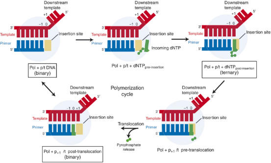
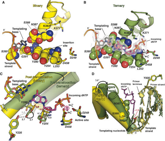
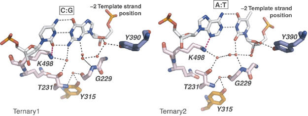
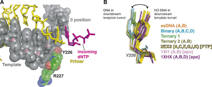
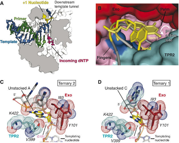
Replicative DNA polymerases (DNAPs) move along template DNA in a processive manner. The structural basis of the mechanism of translocation has been better studied in the A-family of polymerases than in the B-family of replicative polymerases. To address this issue, we have determined the X-ray crystal structures of phi29 DNAP, a member of the protein-primed subgroup of the B-family of polymerases, complexed with primer-template DNA in the presence or absence of the incoming nucleoside triphosphate, the pre- and post-translocated states, respectively. Comparison of these structures reveals a mechanism of translocation that appears to be facilitated by the coordinated movement of two conserved tyrosine residues into the insertion site. This differs from the mechanism employed by the A-family polymerases, in which a conserved tyrosine moves into the templating and insertion sites during the translocation step. Polymerases from the two families also interact with downstream single-stranded template DNA in very different ways.


Figures

Figure 1
Figure 1
The polymerization cycle. Polymerase (pale blue circle) binds a primer-template substrate (blue and red) and then the incoming dNTP (green). In some polymerases, the incoming dNTP binds a pre-insertion site before binding the insertion site (yellow) opposite the templating nucleotide. The polymerase then catalyzes the incorporation of the dNTP into the primer strand, resulting in a primer extended by one nucleotide and a molecule of pyrophosphate bound near the active site. Release of the pyrophosphate is associated with translocation of the new primer terminus out of the insertion site and into the priming site (Yin and Steitz, 2004). Boxes indicate the states captured in our crystal structures. For consistency, template strand numbering refers to the base positions in the initial binary complex.
Figure 2
Figure 2
Comparison of the binary and ternary complexes. The binary complex is shown in yellow and the ternary complex in green. Metals are indicated as magenta spheres. The incoming dNTP from the ternary complex is shown as magenta sticks. The fingers subdomain rotates 14° in going from the opened binary complex to the closed ternary complex. (A) Binary complex. The residues that form the nascent base pair-binding site in the ternary complex are shown as spheres and the active site carboxylates are shown as sticks. The fingers subdomain is shown in cartoon representation. Two conserved tyrosine residues occupy the insertion site. (B) Ternary complex. The conserved lysine residues that interact with the phosphates are also shown. The density from a simulating annealing omit map using phases calculated from a model with the nascent-base pair omitted and amplitudes from the ternary2 data contoured at 2.5 σ is shown as gray mesh for the nascent base pair. (C) Comparison of the binary and ternary complex structures. All of the mechanistically significant amino-acid movements are indicated. Black dashed lines represent interactions. Red dashed lines indicate steric clashes. The distances indicated are in Å. The density shown for metal ion B (manganese) is from an anomalous difference Fourier map calculated using data from 50.0–2.03 Å resolution and contoured at 6 σ. (D) The propagated shift of the DNA base pair planes between the binary and ternary complexes. When the fingers close, residues S388 and N387 (shown as spheres) stack against the templating nucleotide and incoming dNTP, respectively, completing the nascent base pair-binding pocket. Y500 interacts with the phosphate moiety of the priming nucleotide.
Figure 3
Figure 3
Water-mediated interactions maintain sequence nonspecific binding. The C:G base pair is from the ternary1 complex, and the A:T base pair is from the ternary2 complex. Red spheres are water molecules and black dashes are hydrogen bonds. Amino acids are colored by subdomain as in Kamtekar et al (2004).
Figure 4
Figure 4
The I/YxGG/A motif. (A) The primer and template strands from the ternary complex are shown as yellow and gray sticks, respectively. The template strand and the residues of the I/YxGG/A motif are shown as spheres. (B) The two distinct populations of Y226 are shown in sticks based on a superposition of the palm subdomain. The residues are colored by crystal structure.
Figure 5
Figure 5
ssDNA in the downstream template tunnel. (A) A space filling representation of polymerase sliced through a plane showing the primer-template substrate and the single-stranded 5′ template overhang in the downstream template tunnel. The +1 nucleotide is shown in yellow, the incoming nucleotide in magenta, the primer strand in green and the template strand in blue. (B–D) The ssDNA substrate is gray, the +1 nucleotide is yellow. Polymerase residues are colored by domain and subdomain as in Kamtekar et al (2004). (B) An overlay of the substrates from the ternary1 (light yellow and light gray) and ternary2 (dark yellow and dark gray) crystal forms. The polymerase shown is a space filling representation from the ternary1 crystal form. The purine base has no clashes with the protein from the complex containing an unstacked pyrimidine, indicating that the downstream template tunnel does not constrict around pyrimidine bases. (C) A purine base in the unstacked position interacts with residues from the exonuclease domain and the TPR2 subdomain in the ternary2 crystal form. The van der Waals radii of the residues are indicated by dots. (D) A pyrimidine base in the +1 template position in the ternary1 crystal form.
Figure 6
Figure 6
The kink between the +1 and templating nucleotides is a common feature in A- and B-family polymerases. The structures of the B. stearothermophilus DNAPs are from 1L3S (binary) and 1LV5 (ternary). For consistency, all numbers refer to the templating positions before nucleotide addition. In all panels, the template nucleotide of the nascent base pair is shown in orange (−1), the next templating nucleotide in pink (0), and the one 5′ to it in yellow (+1). All residues are shown as spheres, except for the conserved A-family tyrosine (714 in B. stearothermophilus DNAP), which is shown as sticks inside spheres to emphasize its movement, and Y101 from phi29 DNAP shown as sticks inside spheres for clarity. (A) When the fingers are opened in a B-family DNAP, the +1 nucleotide is stabilized by nonconserved hydrophobic interactions, and the templating site is occupied by the templating nucleotide from the last round of incorporation. When the fingers close, no significant movements within the DNA occur. (B) A superposition of the DNA from the binary and ternary complexes of phi29 DNAP. The DNA from the binary complex is colored in lighter colors, whereas that of the ternary complex is in darker colors. There is little difference in the overall positioning of the bases. (C) In the A-family, when the fingers are opened, a conserved tyrosine residue occupies the insertion site. The next templating nucleotide is stabilized in a pre-insertion site of hydrophobic residues from the base of the fingers. When the fingers close, the tyrosine moves out of the templating site, and the templating base moves into the templating site, stacking on the template strand of the upstream duplex. (D) A superposition of the DNA from the binary and ternary complexes of B. stearothermophilus DNAP. The DNA is colored as in (B). The nucleotide in the 0 position is in a very different location in the two complexes.

References

    1. Astatke M, Ng K, Grindley ND, Joyce CM (1998) A single side chain prevents Escherichia coli DNA polymerase I (Klenow fragment) from incorporating ribonucleotides. Proc Natl Acad Sci USA 95: 3402–3407 - PMC - PubMed
    1. Beese LS, Friedman JM, Steitz TA (1993) Crystal structures of the Klenow fragment of DNA polymerase I complexed with deoxynucleoside triphosphate and pyrophosphate. Biochemistry 32: 14095–14101 - PubMed
    1. Blanco L, Salas M (1996) Relating structure to function in phi29 DNA polymerase. J Biol Chem 271: 8509–8512 - PubMed
    1. Blasco MA, Lázaro JM, Bernad A, Blanco L, Salas M (1992) Phi 29 DNA polymerase active site. Mutants in conserved residues Tyr254 and Tyr390 are affected in dNTP binding. J Biol Chem 267: 19427–19434 - PubMed
    1. Blasco MA, Lázaro JM, Blanco L, Salas M (1993) Phi 29 DNA polymerase active site. The conserved amino acid motif ‘Kx3NSxYG' is involved in template-primer binding and dNTP selection. J Biol Chem 268: 16763–16770 - PubMed

Publication types