Review
. 2007 Jul 4;2(7):e583.
doi: 10.1371/journal.pone.0000583.

Immunodominance of lytic cycle antigens in Epstein-Barr virus-specific CD4+ T cell preparations for therapy

Affiliations
Review

Immunodominance of lytic cycle antigens in Epstein-Barr virus-specific CD4+ T cell preparations for therapy

Dinesh Adhikary et al. PLoS One. .

Abstract

Background: Epstein-Barr virus (EBV) is associated with a number of human malignancies. EBV-positive post-transplant lymphoproliferative disease in solid organ and hematopoietic stem cell transplant recipients has been successfully treated by the adoptive transfer of polyclonal EBV-specific T cell lines containing CD4+ and CD8+ T cell components. Although patients receiving T cell preparations with a higher CD4+ T cell proportion show better clinical responses, the specificity of the infused CD4+ component has remained completely unknown.

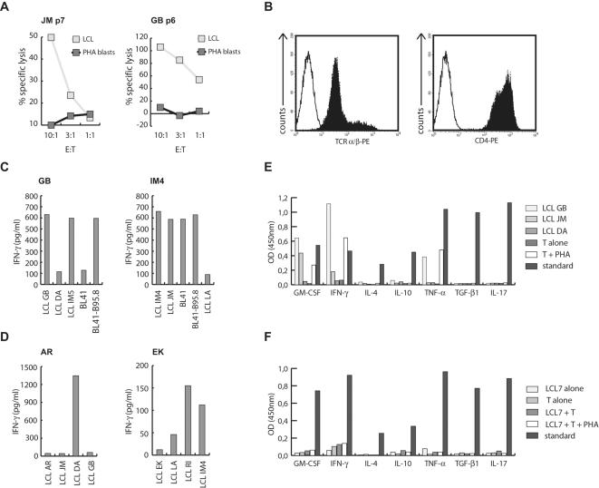
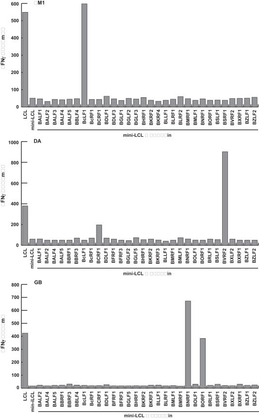
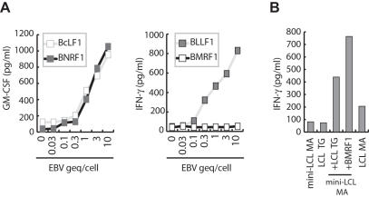
Methodology/principal findings: We generated LCL-stimulated T cell lines from 21 donors according to clinical protocols, and analyzed the antigen specificity of the CD4+ component in EBV-specific T cell preparations using a genetically engineered EBV mutant that is unable to enter the lytic cycle, and recombinantly expressed and purified EBV proteins. Surprisingly, CD4+ T cell lines from acutely and persistently EBV-infected donors consistently responded against EBV lytic cycle antigens and autoantigens, but barely against latent cycle antigens of EBV hitherto considered principal immunotherapeutic targets. Lytic cycle antigens were predominantly derived from structural proteins of the virus presented on MHC II via receptor-mediated uptake of released viral particles, but also included abundant infected cell proteins whose presentation involved intercellular protein transfer. Importantly, presentation of virion antigens was severely impaired by acyclovir treatment of stimulator cells, as currently performed in most clinical protocols.

Conclusions/significance: These results indicate that structural antigens of EBV are the immunodominant targets of CD4+ T cells in LCL-stimulated T cell preparations. These findings add to our understanding of the immune response against this human tumor-virus and have important implications for the improvement of immunotherapeutic strategies against EBV.


Conflict of interest statement

Competing Interests: The authors have declared that no competing interests exist.

Figures

Figure 1
Figure 1. Generation and characterization of LCL-stimulated CD4+ T cell lines.
T cell lines established from EBV-positive donors by LCL stimulation lysed autologous LCL but not PHA blasts after 4–8 passages at different effector-to-target (E:T) ratios. (B) FACS analysis of CD4+ cell lines established from LCL-stimulated bulk T cell lines by magnetic sorting demonstrated that >95% of the cells were TCRα/β+ and CD4+. (C) As demonstrated for donor GB, all TH cell lines established from healthy virus carriers responded against autologous and MHCII-matched allogeneic LCL, as well as MHCII-matched EBV-positive (BL41-B95.8) but not EBV-negative (BL41) Burkitt's lymphoma cell lines. TH cell lines from IM patients showed similar responses against autologous LCL, but as exemplified by the T cell line from IM4, some of these lines also recognized EBV-negative BL cell lines. (D) LCL-stimulated TH cell lines from EBV-negative donors showed minimal if any responses against autologous LCL, but vigorous responses against some allogeneic targets. (E) EBV-reactive TH cell lines secreted GM-CSF, IFN-γ, and TNF-α, but not IL-4, IL-10, IL-17, or TGF-β1 in response to stimulation with autologous (GB) or MHCII-matched allogeneic (JM) LCL, or non-specific activation by PHA. The MHC-mismatched LCL DA served as negative control. The following standards were included: GM-CSF: 1,900 pg/ml; IFN-γ: pg/ml; IL-4: 250 pg/ml; IL-10: 2,100 pg/ml; TNF-α: 2,900 pg/ml; TGF-β1: 1,450 pg/ml; IL-17: 1,700 pg/ml. (F) The T cell line IM7 displayed a novel “non-responder” phenotype. This T cell line proliferated in response to stimulation with autologous LCL and IL-2, but failed to secrete any of the indicated cytokines even after stimulation with autologous LCL plus PHA.
Figure 2
Figure 2. Latent cycle antigens of EBV are not the principal targets of LCL-stimulated TH cells.
EBV-specific TH cell lines from different donors at different passages were tested for recognition of autologous PBMC pulsed separately with the eight antigenically distinct latent cycle proteins of the virus. Except for the T cell lines from donors DA and MS, which showed weak responses against EBNA3C, neither early nor late passage TH cell lines responded against EBV latent cycle proteins.
Figure 3
Figure 3. EBV-reactive TH cell lines recognize autologous LCL but not mini-LCL.
LCL-stimulated TH cell lines showing EBV reactivity were tested for recognition of autologous LCL and mini-LCL established by infection of B cells with an EBV mutant unable to enter the lytic cycle. After three to twenty passages, mini-LCL reactivity of all TH cell lines had dropped to background levels while responses against LCL were maintained even after extended periods of in vitro culture. (B) Responses against LCL and mini-LCL of different passage TH cell lines were assessed by IFN-γ ELISPOT. With the exception of the T cell line from SM, early passage TH cell lines from healthy virus carriers recognized LCL and mini-LCL, but responses against mini-LCL disappeared with further rounds of stimulation. By contrast, early and late passage T cell lines from IM3, which had failed to show EBV-reactivity in earlier experiments, responded similarly against both types of target cells. SFC, spot forming cells.
Figure 4
Figure 4. EBV-reactive TH cell lines target lytic cycle antigens of the virus.
The antigens recognized by TH cell lines that responded against LCL but had lost mini-LCL reactivity were identified using mini-LCL pulsed with recombinant EBV proteins. Responses of three representative CD4+ T cell lines (IM1, DA, and GB) against 30 different lytic cycle proteins are shown.
Figure 5
Figure 5. Lytic antigens are transferred between cells by virions and released proteins.
CD4+ T cells specific for BLLF1, BMRF1, BcLF1 or BNRF1 were tested for recognition of mini-LCL pulsed with purified viral particles. Whereas BcLF1, BLLF1, and BNRF1-specific T cells responded against mini-LCL pulsed with less than 1 genome equivalent (geq) of the virus/cell, BMRF1-specific T cells failed to recognize mini-LCL pulsed with much higher doses of virus. (B) To detect transfer of antigen between cells, BMRF1-specific T cells were tested for recognition of mini-LCL, MHC-mismatched LCL, and the mix of these two lines. While neither line alone was recognized by the T cells, 24 hours of co-culture sensitized the cell mix for recognition.
Figure 6
Figure 6. Presentation of virion antigens is impaired in acyclovir-treated LCL.
LCL either left untreated or treated with acyclovir for two weeks were used as targets for BMRF1 (A), autoantigen (B), or BNRF1-specific T cells (C). Acyclovir treatment neither affected presentation of the autoantigen nor the EBV early lytic cycle antigen BMRF1, but severely reduced the presentation of the virion antigen BNRF1. (D) T cell lines generated by repeated stimulation of peripheral blood CD4+ cells with acyclovir-treated LCL recognized LCL and mini-LCL that had been pulsed with purified EBV particles, suggesting that late lytic cycle antigen-specific T cells still expand under these stimulation conditions.

Similar articles

Cited by

References

    1. Kuppers R. B cells under influence: transformation of B cells by Epstein-Barr virus. Nat Rev Immunol. 2003;3:801–812. - PubMed
    1. Young LS, Rickinson AB. Epstein-Barr virus: 40 years on. Nat Rev Cancer. 2004;4:757–768. - PubMed
    1. Rickinson AB, Kieff E. Epstein-Barr virus. In: Knipe DM, Howley PM, editors. Field's Virology. 5th ed. Philadelphia: Lippincott-Raven; 2006. pp. 2655–2700.
    1. Papesch M, Watkins R. Epstein-Barr virus infectious mononucleosis. Clin Otolaryngol Allied Sci. 2001;26:3–8. - PubMed
    1. Thorley-Lawson DA, Gross A. Persistence of the Epstein-Barr virus and the origins of associated lymphomas. N Engl J Med. 2004;350:1328–1337. - PubMed

MeSH terms