Review
. 2007 Dec;455(3):375-96.
doi: 10.1007/s00424-007-0296-1. Epub 2007 Jul 5.

Mitochondria and Ca(2+) signaling: old guests, new functions

Affiliations
Review

Mitochondria and Ca(2+) signaling: old guests, new functions

Wolfgang F Graier et al. Pflugers Arch. 2007 Dec.

Abstract

Mitochondria are ancient endosymbiotic guests that joined the cells in the evolution of complex life. While the unique ability of mitochondria to produce adenosine triphosphate (ATP) and their contribution to cellular nutrition metabolism received condign attention, our understanding of the organelle's contribution to Ca(2+) homeostasis was restricted to serve as passive Ca(2+) sinks that accumulate Ca(2+) along the organelle's negative membrane potential. This paradigm has changed radically. Nowadays, mitochondria are known to respond to environmental Ca(2+) and to contribute actively to the regulation of spatial and temporal patterns of intracellular Ca(2+) signaling. Accordingly, mitochondria contribute to many signal transduction pathways and are actively involved in the maintenance of capacitative Ca(2+) entry, the accomplishment of Ca(2+) refilling of the endoplasmic reticulum and Ca(2+)-dependent protein folding. Mitochondrial Ca(2+) homeostasis is complex and regulated by numerous, so far, genetically unidentified Ca(2+) channels, pumps and exchangers that concertedly accomplish the organelle's Ca(2+) demand. Notably, mitochondrial Ca(2+) homeostasis and functions are crucially influenced by the organelle's structural organization and motility that, in turn, is controlled by matrix/cytosolic Ca(2+). This review intends to provide a condensed overview on the molecular mechanisms of mitochondrial Ca(2+) homeostasis (uptake, buffering and storage, extrusion), its modulation by other ions, kinases and small molecules, and its contribution to cellular processes as fundamental basis for the organelle's contribution to signaling pathways. Hence, emphasis is given to the structure-to-function and mobility-to-function relationship of the mitochondria and, thereby, bridging our most recent knowledge on mitochondria with the best-established mitochondrial function: metabolism and ATP production.


Figures
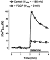
Fig. 1
Fig. 1
Effect of membrane depolarization of the mitochondria on the organelle’s Ca2+ sequestration upon cell stimulation with the IP3-generating agonist histamine. Mitochondrial Ca2+ signaling was measured in single endothelial cells that expressed mitochondrial targeted ratiometric pericam using a high resolution fluorescence microscope as described previously [127, 128]. In Ca2+ containing solution, mitochondria were depolarized by 2 μM FCCP. Changes of the fluorescence intensity at 430 nm excitation and 535 nm emission are shown in percent of the maximal effect of histamine under control conditions (i.e., in the absence of the chemical uncoupler)
Fig. 2
Fig. 2
Effect of membrane depolarization of the mitochondria on the organelle’s morphology and structural integrity. The architectural organization of mitochondria was visualized in human endothelial cells, which transiently expressed mitochondrial-targeted DsRed using an array confocal laser scanning microscope as described previously [160, 215]. a Under basal conditions, mitochondria consist as tubular, highly interconnected network. b After treatment with 2 μM FCCP for 10 min, mitochondria fragment and form singular round mitochondria. c Time course of FCCP induced fragmentation of tubular mitochondria. Upon membrane depolarization by the chemical uncoupler, mitochondrial fragmentation is preluded by the formation of ring-like structures
Fig. 3
Fig. 3
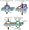
Schematic illustration of putative pathways for mitochondrial Ca2+ fluxes across the inner mitochondrial membrane (IMM; a–b ruthenium red-sensitive MCU). a The ruthenium red-sensitive MCU might consist of just one type of protein (s). Probably UCP2/UCP3 or some other, so far unidentified, protein works alone or forms homomultimeres to establish MCU. However, because of our recent work, this possibility seems unlikely at least for UCP2/UCP3 [215]. b Alternatively, MCU is accomplished by the assembly of a multi protein complex that may form a Ca2+ permeable channel similar to what has been described for the mitochondrial permeability transition pore. Our recent work favors this possibility and points to a fundamental contribution of UCP2/UCP3 in this process [215]. c The ruthenium red-insensitive mitochondrial Ca2+ uptake pathways that either depend on ATP or other ions (Na+, H+) might also contribute to mitochondrial Ca2+ signaling under certain conditions and tissues
Fig. 4
Fig. 4
Correlation between the effect of an inhibition of SERCA on the cytosolic and mitochondrial Ca2+ concentration. Cytosolic free Ca2+ concentration ([Ca2+]cyto) was measured in single human endothelial cells using fura-2 in a high resolution fluorescence microscope as described previously [73, 74, 125]. Analog experiments were performed with cells, which stably expressed mitochondrial targeted ratiometric pericam, to monitor mitochondrial free Ca2+ content ([Ca2+]mito). Changes in [Ca2+]mito are expressed as 1-F430/F0, as at 430 nm excitation the fluorescence intensity reflects the Ca2+ sensitivity of this sensor [128]. As indicated, SERCA was inhibited by the addition of 2,5-di-tert-butylhydroquinone (BHQ, 15 μM)
Fig. 5
Fig. 5
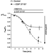
Effect of an inhibition of the mitochondrial Na+/Ca2+ exchanger on histamine-induced changes in the pH of the mitochondrial matrix. Endothelial cells, which stably express mitochondrial targeted ratiometric pericam were used to monitor changes of the matrix pH by following the pH sensitive wavelength from the sensor. Cells were illuminated at 480 nm and emission was collected at 535 nm on a high-resolution fluorescence microscope [127, 128]. As indicated cells were stimulated with 100 μM histamine in the absence (continous line, open circles) or in the presence of 20 μM CGP 37157 (dotted line, filled circles)
Fig. 6
Fig. 6
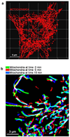
Mitochondrial morphology and motility. a Highly interconnected mitochondria in a single living HeLa cell that transiently expressed mitochondria-targeted DsRed. Z-Scans were performed on an array confocal laser scanning microscope applying 514 nm excitation and measuring 570 nm emission [160]. 3-D reconstruction of the mitochondrial network was performed using Imaris 4.2 software. b Overlay of mitochondrial structures in a single endothelial cell expressing mitochondria-targeted DsRed at 0, 5 and 10 min

Similar articles

Cited by

References

    1. Alirol E, Martinou JC. Mitochondria and cancer: is there a morphological connection? Oncogene. 2006;25:4706–4716. - PubMed
    1. Andreyev AY, Kushnareva YE, Starkov AA. Mitochondrial metabolism of reactive oxygen species. Biochemistry (Mosc) 2005;70:200–214. - PubMed
    1. Ardehali H, Chen Z, Ko Y, Mejia-Alvarez R, Marban E. Multiprotein complex containing succinate dehydrogenase confers mitochondrial ATP-sensitive K+ channel activity. Proc Natl Acad Sci USA. 2004;101:11880–11885. - PMC - PubMed
    1. Armstrong JS. The role of the mitochondrial permeability transition in cell death. Mitochondrion. 2006;6:225–234. - PubMed
    1. Arnaudeau S, Kelley WL, Walsh JV, Jr, Demaurex N. Mitochondria recycle Ca2+ to the endoplasmic reticulum and prevent the depletion of neighboring endoplasmic reticulum regions. J Biol Chem. 2001;276:29430–29439. - PubMed

Publication types

MeSH terms

LinkOut - more resources