. 2007;9(4):R65.
doi: 10.1186/ar2223.

Expression and function of junctional adhesion molecule-C in human and experimental arthritis

Affiliations

Expression and function of junctional adhesion molecule-C in human and experimental arthritis

Gaby Palmer et al. Arthritis Res Ther. 2007.

Abstract

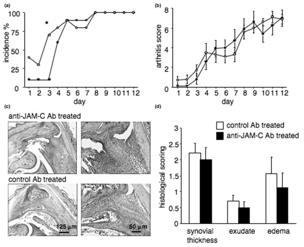
Junctional adhesion molecule-C (JAM-C) is an adhesion molecule involved in transendothelial migration of leukocytes. In this study, we examined JAM-C expression in the synovium and investigated the role of this molecule in two experimental mouse models of arthritis. JAM-C expression was investigated by reverse transcriptase-polymerase chain reaction and immunohistochemistry. The effects of a monoclonal anti-JAM-C antibody were assessed in antigen-induced arthritis (AIA) and K/BxN serum transfer-induced arthritis. JAM-C was expressed by synovial fibroblasts in the lining layer and associated with vessels in the sublining layer in human and mouse arthritic synovial tissue. In human tissue, JAM-C expression was increased in rheumatoid arthritis (RA) as compared to osteoarthritis synovial samples (12.7 +/- 1.3 arbitrary units in RA versus 3.3 +/- 1.1 in OA; p < 0.05). Treatment of mice with a monoclonal anti-JAM-C antibody decreased the severity of AIA. Neutrophil infiltration into inflamed joints was selectively reduced as compared to T-lymphocyte and macrophage infiltration (0.8 +/- 0.3 arbitrary units in anti-JAM-C-treated versus 2.3 +/- 0.6 in isotype-matched control antibody-treated mice; p < 0.05). Circulating levels of the acute-phase protein serum amyloid A as well as antigen-specific and concanavalin A-induced spleen T-cell responses were significantly decreased in anti-JAM-C antibody-treated mice. In the serum transfer-induced arthritis model, treatment with the anti-JAM-C antibody delayed the onset of arthritis. JAM-C is highly expressed by synovial fibroblasts in RA. Treatment of mice with an anti-JAM-C antibody significantly reduced the severity of AIA and delayed the onset of serum transfer-induced arthritis, suggesting a role for JAM-C in the pathogenesis of arthritis.


Figures

Figure 1
Figure 1
Expression of junctional adhesion molecule-C (JAM-C) in human arthritic synovium. Expression of JAM-C was examined by immunohistochemistry in human osteoarthritis (OA) (a) and rheumatoid arthritis (RA) (b) synovial tissue. The panels labeled 1 show the original magnification (× 100). The panels labeled 2 show expression of JAM-C in the synovial lining layer (magnification × 400), and panels labeled 3 show JAM-C associated with blood vessels in the sublining (magnification × 400). The panels labeled 4 show negative control sections incubated with preimmune serum. Scale bars = 60 μm (panels 1 and 4) and 15 μm (panels 2 and 3). (c) Quantification of JAM-C expression in OA and RA synovial tissues. JAM-C immunohistochemical synovial tissue sections from four different OA and RA patients were scanned, and the surface of immunoreactive areas was determined and expressed as the percentage of the surface of the image examined. Results are expressed as the mean ± standard error of the mean. *p < 0.001 OA versus RA as assessed using the Wilcoxon rank sum test. (d) Reverse transcriptase-polymerase chain reaction (PCR) analysis of JAM-C mRNA expression in human synovial tissue samples and in cultured human synovial fibroblasts. A representative agarose gel electrophoresis of PCR products is shown. bp, base pairs; H2O, polymerase chain reaction negative control; hSF1 and hSF2, human rheumatoid arthritis synovial fibroblast cultures from two different patients used after the third passage; OA, osteoarthritis synovial tissue; RA, rheumatoid arthritis synovial tissue; RT neg, non-reverse-transcribed sample.
Figure 2
Figure 2
Expression of junctional adhesion molecule-C (JAM-C) in mouse arthritic synovium and synovial fibroblasts. Expression of JAM-C was examined by immunohistochemistry in knee joints of mice with antigen-induced arthritis (AIA) (a) and in paws of mice with K/BxN serum transfer-induced arthritis (b). The left panels show expression of JAM-C in the synovial lining layer (upper panel) and associated with blood vessels in the sublining (lower panel). The right panels show negative control sections incubated with preimmune serum. Original magnification × 400 (scale bar = 20 μm). (c) Reverse transcriptase-polymerase chain reaction (PCR) analysis of JAM-C mRNA expression in mouse synovial tissue samples and in cultured mouse synovial fibroblasts. A representative agarose gel electrophoresis of PCR products is shown. AIA1 and AIA2, antigen-induced arthritis synovial tissue; bp, base pairs; H2O, polymerase chain reaction negative control; K/BxN1 and K/BxN2, K/BxN serum transfer-induced arthritis synovial tissue; mSF1 and mSF2, two different mouse synovial fibroblast cultures isolated from antigen-induced arthritis synovial tissue; RTneg, non reverse-transcribed sample.
Figure 3
Figure 3
Treatment with anti-JAM-C antibody decreases the severity of antigen-induced arthritis. (a) Joint inflammation on days 1, 3, and 7 after intra-articular methylated bovine serum albumin (mBSA) injection. Results are expressed as the ratio of technetium-99m (99mTc) uptake in the arthritic over the non-inflamed knee. The mean ± standard error of the mean (SEM) of the ratios is shown for anti-JAM-C antibody-treated (n = 10 for days 1 and 3, n = 6 for day 7; black symbols) and isotype-matched control antibody-treated (n = 10 for days 1 and 3, n = 5 for day 7; open symbols) mice. Joint inflammation was significantly reduced in anti-JAM-C antibody-treated mice on day 3. (b) Representative histological sections for anti-JAM-C antibody-treated (upper panel) and isotype-matched control antibody-treated (lower panel) mice 8 days after intra-articular mBSA injection (original magnification × 40, scale bar = 125 μm). Arrowheads: cartilage erosions; asterisks: pannus; broken lines: synovial thickness. (c) Histological scores shown as the mean ± SEM for anti-JAM-C antibody-treated (n = 6, black columns) and isotype-matched control antibody-treated (n = 5, open columns) mice 8 days after intra-articular mBSA injection. Synovial inflammation was significantly reduced in anti-JAM-C antibody-treated mice. (d) Circulating levels of serum amyloid A (SAA) on days 4 and 8 after intra-articular mBSA injection. Results shown represent the mean ± SEM for anti-JAM-C antibody-treated (n = 5 on day 4, n = 6 on day 8, black columns) and isotype-matched control antibody-treated (n = 5 at both time points, open columns) mice. Serum SAA levels were significantly decreased in anti-JAM-C antibody-treated mice on day 4. *p < 0.05 versus control mice, as assessed by analysis of variance. Ab, antibody; JAM-C, junctional adhesion molecule-C.
Figure 4
Figure 4
Treatment with anti-JAM-C antibody decreases the T-cell response. (a) Proliferation of spleen cells isolated from anti-JAM-C antibody-treated (n = 5) and isotype-matched control antibody-treated (n = 5) mice. Cells were restimulated in vitro with methylated bovine serum albumin (mBSA) (10 μg/ml, hatched columns) or concanavalin A (ConA) (5 μg/ml) or were left unstimulated (white columns). Results, expressed as fold increase in proliferation of stimulated over unstimulated cultures, represent the mean ± standard error of the mean (SEM) for each group of mice. Proliferation was significantly increased by stimulation with mBSA or ConA in cells isolated from isotype-matched control antibody-treated mice; *p < 0.05 versus unstimulated cultures. This proliferative response to mBSA and ConA was significantly reduced in cells of anti-JAM-C antibody-treated mice as compared to cells isolated from isotype-matched control antibody-treated mice; &p < 0.05 versus cells isolated from isotype-matched control antibody-treated mice with similar stimulation, as assessed by analysis of variance (ANOVA). (b) Interferon-gamma (IFN-γ) production by spleen cells isolated from anti-JAM-C antibody-treated (n = 5) and isotype-matched control antibody-treated (n = 5) mice. Cells were restimulated in vitro with mBSA (10 μg/ml, hatched columns) or ConA (5 μg/ml) or were left unstimulated (white columns). Results shown represent the mean ± SEM for each group of mice. IFN-γ production was significantly increased by stimulation with mBSA or ConA in cells isolated from isotype-matched control antibody-treated mice; *p < 0.05 versus unstimulated cultures. This IFN-γ response to mBSA and ConA was markedly reduced in cells of anti-JAM-C antibody-treated mice as compared to cells isolated from isotype-matched control antibody-treated mice; &p < 0.05 versus cells isolated from isotype-matched control antibody-treated mice with similar stimulation, as assessed by ANOVA. Serum levels of anti-mBSA total IgG (c), IgG1 ([d], left panel), and IgG2a ([d], right panel) on days 4 and 8 after intra-articular mBSA injection. Results shown represent the mean ± SEM for anti-JAM-C antibody-treated (n = 6, black columns) and isotype-matched control antibody-treated (n = 5, open columns) mice. *p < 0.05 versus control mice, as assessed by ANOVA. Ab, antibody; A.U., arbitrary units; Ig = immunoglobulin; JAM-C, junctional adhesion molecule-C; O.D., optical density.
Figure 5
Figure 5
Treatment with anti-JAM-C antibody decreases neutrophil infiltration into the joints during antigen-induced arthritis. Infiltration of neutrophils (a), lymphocytes (b), and macrophages (c) into the synovium was detected by immunohistochemistry using anti-MPO, anti-CD3, and anti-MAC-2 antibodies, respectively. The left panels show representative knee joint sections of control and anti-JAM-C antibody-treated mice. Original magnification × 400 (scale bar = 25 μm). In the right panels, leukocyte infiltration per field was evaluated by semi-quantitative scoring for anti-JAM-C antibody-treated (n = 6, black columns) and isotype-matched control antibody-treated (n = 5, open columns) mice. There was a significant decrease in synovial neutrophil infiltration in anti-JAM-C antibody-treated mice as compared to isotype-matched antibody-treated controls. *p < 0.05 versus control mice, as assessed by analysis of variance. Ab, antibody; JAM-C, junctional adhesion molecule-C.
Figure 6
Figure 6
Treatment with anti-JAM-C antibody delays onset of K/BxN serum transfer-induced arthritis. (a) Incidence of K/BxN serum transfer-induced arthritis is shown for anti-JAM-C antibody-treated (n = 10, black symbols) and isotype-matched control antibody-treated (n = 10, open symbols) mice. Results are expressed as the percentage of arthritic mice per group. Incidence of arthritis was significantly lower in anti-JAM-C antibody-treated mice as compared to isotype-matched antibody-treated controls on day 3. *p < 0.05 versus control mice, as assessed by chi-square test. (b) Severity of K/BxN serum transfer-induced arthritis. Arthritis was evaluated by clinical assessment of arthritis severity scores for anti-JAM-C antibody-treated (n = 10, black symbols) and isotype-matched control antibody-treated (n = 10, open symbols) mice. Results shown represent the mean ± standard error of the mean (SEM) for each group of mice. There were no significant differences between the groups. (c) Histological assessment of arthritis. Representative sections are shown for anti-JAM-C antibody-treated (upper panels) and isotype-matched control antibody-treated (lower panels) mice 13 days after serum transfer. Original magnifications × 40 (left panel) (scale bar = 125 μm) and × 100 (right panel) (scale bar = 50 μm). (d) Histological scores for synovial thickness, exudates, and edema 13 days after serum transfer. Results shown represent the mean ± SEM for anti-JAM-C antibody-treated (n = 10, black columns) and isotype-matched control antibody-treated (n = 10, open columns) mice. No significant differences were observed between the groups. Ab, antibody; JAM-C, junctional adhesion molecule-C.

Similar articles

Cited by

References

    1. Butcher EC. Leukocyte-endothelial cell recognition: three (or more) steps to specificity and diversity. Cell. 1991;67:1033–1036. doi: 10.1016/0092-8674(91)90279-8. - DOI - PubMed
    1. Springer TA. Traffic signals for lymphocyte recirculation and leukocyte emigration: the multistep paradigm. Cell. 1994;76:301–314. doi: 10.1016/0092-8674(94)90337-9. - DOI - PubMed
    1. Schenkel AR, Mamdouh Z, Chen X, Liebman RM, Muller WA. CD99 plays a major role in the migration of monocytes through endothelial junctions. Nat Immunol. 2002;3:143–150. doi: 10.1038/ni749. - DOI - PubMed
    1. Muller WA, Weigl SA, Deng X, Phillips DM. PECAM-1 is required for transendothelial migration of leukocytes. J Exp Med. 1993;178:449–460. doi: 10.1084/jem.178.2.449. - DOI - PMC - PubMed
    1. Johnson-Leger CA, Aurrand-Lions M, Beltraminelli N, Fasel N, Imhof BA. Junctional adhesion molecule-2 (JAM-2) promotes lymphocyte transendothelial migration. Blood. 2002;100:2479–2486. doi: 10.1182/blood-2001-11-0098. - DOI - PubMed

Publication types

MeSH terms