. 2007 Jul 6:7:109.
doi: 10.1186/1471-2148-7-109.

Molecular evolution of the reactive oxygen-generating NADPH oxidase (Nox/Duox) family of enzymes

Affiliations

Molecular evolution of the reactive oxygen-generating NADPH oxidase (Nox/Duox) family of enzymes

Tsukasa Kawahara et al. BMC Evol Biol. .

Abstract

Background: NADPH-oxidases (Nox) and the related Dual oxidases (Duox) play varied biological and pathological roles via regulated generation of reactive oxygen species (ROS). Members of the Nox/Duox family have been identified in a wide variety of organisms, including mammals, nematodes, fruit fly, green plants, fungi, and slime molds; however, little is known about the molecular evolutionary history of these enzymes.

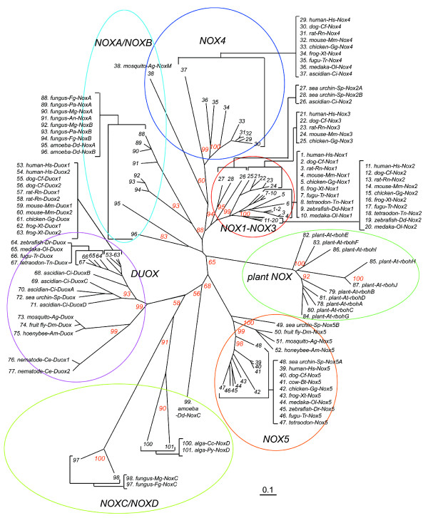
Results: We assembled and analyzed the deduced amino acid sequences of 101 Nox/Duox orthologs from 25 species, including vertebrates, urochordates, echinoderms, insects, nematodes, fungi, slime mold amoeba, alga and plants. In contrast to ROS defense enzymes, such as superoxide dismutase and catalase that are present in prokaryotes, ROS-generating Nox/Duox orthologs only appeared later in evolution. Molecular taxonomy revealed seven distinct subfamilies of Noxes and Duoxes. The calcium-regulated orthologs representing 4 subfamilies diverged early and are the most widely distributed in biology. Subunit-regulated Noxes represent a second major subdivision, and appeared first in fungi and amoeba. Nox5 was lost in rodents, and Nox3, which functions in the inner ear in gravity perception, emerged the most recently, corresponding to full-time adaptation of vertebrates to land. The sea urchin Strongylocentrotus purpuratus possesses the earliest Nox2 co-ortholog of vertebrate Nox1, 2, and 3, while Nox4 first appeared somewhat later in urochordates. Comparison of evolutionary substitution rates demonstrates that Nox2, the regulatory subunits p47phox and p67phox, and Duox are more stringently conserved in vertebrates than other Noxes and Nox regulatory subunits. Amino acid sequence comparisons identified key catalytic or regulatory regions, as 68 residues were highly conserved among all Nox/Duox orthologs, and 14 of these were identical with those mutated in Nox2 in variants of X-linked chronic granulomatous disease. In addition to canonical motifs, the B-loop, TM6-FAD, VXGPFG-motif, and extreme C-terminal regions were identified as important for Nox activity, as verified by mutational analysis. The presence of these non-canonical, but highly conserved regions suggests that all Nox/Duox may possess a common biological function remained in a long history of Nox/Duox evolution.

Conclusion: This report provides the first comprehensive analysis of the evolution and conserved functions of Nox and Duox family members, including identification of conserved amino acid residues. These results provide a guide for future structure-function studies and for understanding the evolution of biological functions of these enzymes.


Figures

Figure 1
Figure 1
Schematic domain structures of Nox family. (A) The Nox domain possesses 6 transmembrane α-helices (I through VI in boxes), two hemes ("heme" in diamonds), and predicted sub-regions that provide binding cavities for a co-enzyme FAD (FAD1 and FAD2) and for a substrate NADPH (N-I, N-II, N-III, and N-IV). (B) All members of Nox/Duox family contain the Nox domains. Abbreviations are: "EF" refers to any domain containing one or more EF-hand motifs; "peroxidase" refers to a region that is homologous to a heme-containing peroxidase; "N" refers to an asparagine-rich region; "4 × TM", four predicted transmembrane α-helical segments; "At-rbohD" is A. thaliana respiratory burst oxidase homolog-D; "Ag" is A. gambiae; "Dd" is D. discoideum; "Mg" is M. grisea; "Cc" is C. crispus.
Figure 2
Figure 2
Molecular taxonomy of the Nox domains of Nox/Duox proteins. Amino acid sequences of the following species were trimmed to the length corresponding to human Nox2 and were aligned: human-Hs, H. sapiens; Cow-Bt, B. Taurus; dog-Cf, C. familliaris; rat-Rn, R. norvegicus; mouse-Mm, M. musculus; opossum-Md, M. domestica; chicken-Gg, G. gallus; frog-Xt, X. tropicalis; zebrafish-Dr, D. rerio; fugu-Tr, T. rubripes; tetraodon-Tn, T. nigroviridis; medaka-Ol, O. latipes; ascidian-Ci, C. intestinalis; sea urchin-Sp, S. purpuratus; fruit fly-Dm, D. melanogaster; mosquito-Ag, A. gambiae; honeybee-Am, A. mellifera; nematode-Ce, C. elegans; plant-At,A. thaliana; amoeba-Dd, D. discoideum; fungus-Pa, P. anserina, fungus-An, A. nidulans; fungus-Mg, M. grisea; fungus-Fg, F. graminearum; alga-Cc, C. crispus; and alga-Py, P. yezoensis. Each subfamily is indicated by a colored circle, and bootstrap values of 1,000 replications are shown at the major branches as percentages. Evolutionary distances (inferior bar) are equivalent to 0.1 amino acid substitution per site.
Figure 3
Figure 3
Syntenies of Nox/Duox genes. (A) Summary of the occurrence of Nox/Duox genes within eukaryotes. The number indicates the orthologs in each organism. Superscripted letters "a" and "b" represent incomplete amino acid sequences predicted from nucleotide fragments: a, 221 amino acid length of T. nigroviridis Nox4 and b, 201 amino acids of D. rerio Nox4 (sequences are shown in SD1). Superscripted letters "c" and "d" indicate these Nox5 orthologs do not contain N-terminal EF-hand-containing domain (#45 and #47 in Figure 2; sequences are shown in Additional file 4). A parenthesis represents an ambiguous classification of the "Ag-NoxM" (#38 in Figure 2) in the NoxA/NoxB subgroup. (B-G) Syntenies of the indicated vertebrate Noxes are shown. Genes are aligned in columns to illustrate orthology. Chromosome (Chr) or scaffold (Sc) numbers are indicated on the right.
Figure 4
Figure 4
Loop and segment sizes joining transmembrane regions and canonical NADPH-oxidase domains. After alignment of canonical and TM domains, the numbers of amino acids linking these regions were counted. I to VI in boxes indicate the six predicted TM α-helices, and FAD-binding subregions (FAD1 and FAD2) and the four NADPH-binding subregions (NADPH1 to NADPH4) are shown. Solid lines show the relative length of loops and linkers that are characteristic of specific Nox subfamilies. The numbers of amino acids are indicated. Broken lines show the average lengths. More detailed information on the number of residues linking each of the canonical regions for each Nox subtype is provided in Additional file 1.
Figure 5
Figure 5
Comparison of molecular clocks of Nox domains.(A) Amino acid sequences were trimmed to the length of human Nox2 as Nox domains, and the numbers of amino acid substitutions per site relative to the human isolog were counted. Each data point represents amino acid substitution rates per site per 109 years for a given isolog (see Additional file 3). (B) Each data point represents amino acid substitution rates of regulatory subunits per site per 109 years. Asterisks indicate significance: p < 0.001 (*) or p < 0.002 (**).
Figure 6
Figure 6
Identification of amino acid residues conserved in all Nox/Duox proteins. The single letter amino acid code is used, and consensus amino acid residues and locations are identified based on the alignment in Additional file 5. In the column labeled "consensus", "a" refers to hydrophobic side-chain amino acids. In the column labeled "location", "TM" refers to TM α-helix; and "TM3-heme" and "TM5-heme" refer to predicted heme-ligating histidine residues. NADPH1-2, NADPH2-3, and NADPH3-4 refer to amino acids that connect each canonical NADPH sub-region. Ag refers to A. gambiae. Point mutations in Nox2, which occur in variants of X-linked CGD, that correspond to the identified conserved amino acids are indicated. Exceptions to consensus residues are indicated by asterisks and are listed in Additional file 11.
Figure 7
Figure 7
Effects of mutations of conserved amino acids on Nox enzymatic activity and formation of the Nox2-p22phox complex. (A) Conserved amino acids (circles) are indicated on a schematic of the Nox domain, and residue numbers corresponding to the human Nox2 protein sequence are keyed in Figure 6. Filled circles indicate known point mutations in individual variants of X-linked CGD. (B) HEK293 cells that constitutively express p22phox were co-transfected with wild type (WT) or the indicated mutations of Nox2 along with p47phox, p67phox, and Rac1(V12G) or with empty vector (mock). Each point mutation of human Nox2 is indicated by the single letter amino acid code. ROS production was measured as described, and the values are presented as mean ± SD (n = 4). These experiments have been repeated three times with similar results. (C) Nox2 and p22phox protein expression was probed by Western blotting (WB) with monoclonal antibodies 54.1 and 44.1, respectively. Proteins were immunoprecipitated (IP) with antibody 54.1 prior to SDS-PAGE. The asterisks indicate IgG heavy chain (* in upper panels) and light chain (** in lower panels). Nox2 protein is expressed as both unglycosylated (65 kDa, immature) and glycosylated (90–100 kDa, mature) forms. p22phox co-immunoprecipitated with Nox2 was seen at 22 kDa. These experiments have been repeated more than three times with similar results.
Figure 8
Figure 8
Sequences of EF-hand motifs in Nox5, Duox, plant Nox, and NoxC. (A) EF-hand containing Nox/Duox family members are listed and demonstrate the widespread occurrence of these Noxes. Parentheses represent the presence of Nox5 ortholog in the T. nigroviridis and D. rerio genomes, which clearly belongs in the Nox5 subgroup (#45 and #47 in Figure 2), but appears to lack the N-terminal EF-hand-containing domain as discussed in the text. (B) Based on alignments shown in Additional files 2 and 8, the arrangement of EF-hand motifs within the calcium-binding domain of calcium-regulated Noxes are shown schematically. EF-I to EF-VI in squares indicate each canonical EF-hand motif. TM-I and TM-II indicate the 1st or 2nd TM segments. Asterisks represent atypical EF-hand motifs that differ from consensus sequences at positions 1, 3, or 12, normally the most conserved positions [63]. HLH, refers to a non-canonical helix-loop-helix predicted structure. (C) A structural homology model of the EF-hand-containing domains of human Nox5 (upper panel) and human Duox1 (lower panel) using a comparative protein modeling method (SWISS-MODEL) and visualized with Deep View Swiss-PDB. The N-terminal region of Nox5 and human Duox1 was calculated using the structure of calcineurin B subunit isoform 1 as a fit template. The fit of the N-terminal region of Duox1 corresponding to the first EF-hand-like motif of Nox5 was not accurate enough to determine the molecular model. The arrow indicates the position of Duox1 corresponding to the 4th EF-hand motif of Nox5 and models as a HLH structure that lacks canonical calcium binding amino acid residues. Side chains of canonical EF-hand motifs are indicated. Conserved sequences among EF-hand regions in Noxes and Duoxes are aligned and compared in Additional file 2.
Figure 9
Figure 9
Occurrence of Noxes in the phylogenetic tree. A schematic phylogeny of organisms was created from genomic information [49, 53, 67, 91]. Branch lengths are not proportional to divergence. "Mammals" in this figure include H. sapiens, C. familliaris, R. norvegicus, and M. musculus. Among fungi, yeast (S. pombe and S. cerevisiae) lacked Noxes, although they did possess a protein with similar structural domains, Fre (see Additional file 9). The asterisk indicates an apparent lack of EF-hand-containing Nox/Duox in two fungi; M. grisea, F. graminearum possess an EF-hand-containing Nox, whereas P. anserina and A. nidulans do not.

Similar articles

Cited by

References

    1. Burdon R. Superoxide and hydrogen peroxide in relation to mammalian cell proliferation. Free Radic Biol Med. 1995;18:775–794. doi: 10.1016/0891-5849(94)00198-S. - DOI - PubMed
    1. Lambeth JD. NOX enzymes and the biology of reactive oxygen. Nature Review Immunology. 2004;4:181–189. doi: 10.1038/nri1312. - DOI - PubMed
    1. Bedard K, Krause KH. The NOX family of ROS-generating NADPH oxidases: physiology and pathophysiology. Physiological Reviews. 2007;87:245–313. doi: 10.1152/physrev.00044.2005. - DOI - PubMed
    1. Carol RJ, Dolan L. The role of reactive oxygen species in cell growth: lessons from root hairs. Journal of Experimental Botany. 2006;57:1829–1834. doi: 10.1093/jxb/erj201. - DOI - PubMed
    1. Nauseef WM. Assembly of the phagocyte NADPH oxidase. Histochemistry and Cell Biology. 2004;122:277–291. doi: 10.1007/s00418-004-0679-8. - DOI - PubMed

Publication types