. 2007 Jul 6;27(1):17-28.
doi: 10.1016/j.molcel.2007.06.004.

Compartmentalized phosphorylation of IAP by protein kinase A regulates cytoprotection

Affiliations

Compartmentalized phosphorylation of IAP by protein kinase A regulates cytoprotection

Takehiko Dohi et al. Mol Cell. .

Abstract

Cell death pathways are likely regulated in specialized subcellular microdomains, but how this occurs is not understood. Here, we show that cyclic AMP-dependent protein kinase A (PKA) phosphorylates the inhibitor of apoptosis (IAP) protein survivin on Ser20 in the cytosol, but not in mitochondria. This phosphorylation event disrupts the binding interface between survivin and its antiapoptotic cofactor, XIAP. Conversely, mitochondrial survivin or a non-PKA phosphorylatable survivin mutant binds XIAP avidly, enhances XIAP stability, synergistically inhibits apoptosis, and accelerates tumor growth, in vivo. Therefore, differential phosphorylation of survivin by PKA in subcellular microdomains regulates tumor cell apoptosis via its interaction with XIAP.


Figures

Figure 1
Figure 1. Phosphorylation of survivin by PKA
A. Sequence analysis. The consensus PKA phosphorylation site and survivin sequences between residues 18-23 or 77-85 are shown. Putative PKA phosphorylation sites on survivin S20 and S81 are indicated with an asterisk. B. PKA phosphorylation of survivin, in vitro. Recombinant wild type (WT) survivin, GST or S20/81A survivin double mutant was incubated in a kinase assay in the presence of PKA and 32P-γATP. Phosphorylated bands were detected by autoradiography. C. PKA phosphorylation of survivin peptides. A synthetic peptide duplicating the survivin sequence P12-G30 with or without S20A or T21A substitutions was incubated with PKA in a kinase assay and radioactive bands were detected by autoradiography. D. Characterization of a survivin phospho-specific antibody to S20. Recombinant WT survivin or PKA phosphorylation-defective S20A survivin was incubated in a PKA kinase assay in the presence of unlabeled phosphate, and analyzed by Western blotting with a S20 phospho-specific antibody (Survivin(S20*)), or an antibody to unmodified survivin (Survivin). Survivin(S20*), Survivin phosphorylated on S20. E. Concentration-dependence. Increasing concentrations of recombinant WT survivin (0.06, 0.3, 0.6, 1.2 μg) were incubated in a PKA kinase assay in the presence of unlabeled phosphate, and analyzed by Western blotting. F. Densitometry. Protein bands from the experiment in E were quantified by densitometry and expressed as ratio in the presence or absence of PKA phosphorylation.
Figure 2
Figure 2. Identification of PKA as a survivin S20 kinase, in vivo
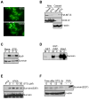
A. Akt kinase assay. Increasing concentrations of recombinant WT survivin (0.06, 0.3, 0.6, 1.2 μg) were incubated in an Akt-1 kinase assay in the presence of unlabeled phosphate, and analyzed by Western blotting. Akt phosphorylation of recombinant Mdm2 (S166) was used as a control. B. Effect of forskolin. INS-1 cells stably expressing WT survivin or S20A survivin (clones #8 and #10) were treated with the PKA activator, forskolin (Fsk) and analyzed by Western blotting. *, non specific. C. Effect of PKI. Top panel, INS-1 cells expressing WT survivin were treated with or without Fsk in the presence or absence of the PKA inhibitor, PKI, and analyzed by Western blotting. Bottom panel, PKA activity in the presence or absence of Fsk. *, non-specific. D. siRNA silencing of PKA. INS-1 cells expressing WT survivin were transfected with non-targeted (control) or PKA-directed siRNA, treated with or without Fsk, and analyzed by Western blotting. For panels A, C, D, a kinase assay with recombinant survivin in the presence or absence of PKA was used as a control.
Figure 3
Figure 3. Differential subcellular phosphorylation of survivin by PKA
A. Subcellular compartmentalization of survivin phosphorylation on S20. MCF-7 cells were fractionated in cytosolic or mitochondrial extracts, and analyzed by Western blotting. A kinase assay with recombinant survivin in the presence or absence of PKA was used as a control. *, non specific. B. Immunoprecipitation. Survivin was immunoprecipitated from cytosolic (left panel) or mitochondrial (right panel) extracts, and pellets or supernatants were analyzed by Western blotting. C. Effect of PP2A. PKA-phosphorylated recombinant survivin was mixed with the indicated increasing concentrations of PP2A, and analyzed by Western blotting. COX-IV was used as a mitochondrial marker. D. Submitochondrial fractionation. Isolated mitochondrial extracts treated with proteinase K were incubated with the indicated increasing concentrations of digitonin, and aliquots of pellets (P) or supernatants (S) were analyzed by Western blotting. E, total cell extracts. *, non specific. Bottom panel, submitochondrial localization of PKA.
Figure 4
Figure 4. Complex formation between mitochondrial survivin and XIAP
A. Mitochondrial discharge of survivin during cell death. INS-1 cells stably expressing mitochondrially-targeted survivin fused to GFP were treated with or without staurosporine (STS), and analyzed by fluorescence microscopy after 8 h. B. Western blotting. INS-1 cells expressing HA-tagged mitochondrially-targeted survivin (HA-MT-S) were treated with STS, and mitochondrial (Mito) or cytosolic extracts were analyzed by Western blotting. C. Immunoprecipitation. Cytosolic extracts from untreated (None) or STS-treated MCF-7 cells were immunoprecipitated with an antibody to survivin, and pellets (P) or supernatants (S) were analyzed by Western blotting. *, non specific. D. In vivo capture assay. Mitochondrial (M) or cytosolic (C) extracts from MCF-7 cells were incubated with GST or GST-XIAP, and bound proteins were analyzed by Western blotting. E. Effect of STS on survivin S20 phosphorylation, in vitro. Recombinant survivin in a PKA kinase assay was incubated with the indicated concentrations of STS, and analyzed by Western blotting. F. Time-course. INS-1 cells stably expressing WT survivin were treated with STS, harvested at the indicated time intervals and analyzed for survivin phosphorylation on S20. A kinase assay with recombinant survivin in the presence or absence of PKA was used as a control.
Figure 5
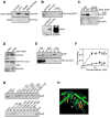
Figure 5. PKA regulation of survivin-XIAP complex
A. Phosphatase treatment. Cytosolic extracts from MCF-7 cells were treated with buffer or calf intestinal phosphatase (CIP), mixed with GST or GST-XIAP, and bound proteins were analyzed by Western blotting. Untreated mitochondrial extracts were used as control. B. Effect of PKA dominant negative (DN) mutant. MCF-7 cells were transfected with pcDNA3 or a PKA-DN cDNA, immunoprecipitated with an antibody to survivin, and immune complexes were analyzed by Western blotting. A kinase assay with recombinant survivin in the presence or absence of PKA was used as a control. Recombinant survivin was used as a control for XIAP binding. C. Differential survivin-XIAP interaction, in vitro. Increasing concentrations of WT survivin or S20E survivin were incubated with GST-XIAP (top panel) or GST (bottom panel), and bound proteins were analyzed by Western blotting. D. In vivo capture assay. Cytosolic extracts from MCF-7 cells transfected with S20A or S20E survivin were incubated with GST-XIAP (top panel) or GST (bottom panel), and bound proteins were analyzed by Western blotting. E. Co-immunoprecipitation. MCF-7 cells expressing HA-tagged S20A (A) or S20E (E) survivin were immunoprecipitated with IgG or an antibody to HA, and the immune complexes were analyzed by Western blotting. F. PKA modulation of survivin-XIAP affinity. WT survivin or S20A survivin was incubated in a PKA kinase assay with unlabeled phosphate, and binding to recombinant XIAP was analyzed by Western blotting. Data are expressed as the ratio between phosphorylated and unphosphorylated survivin binding to XIAP, and are the mean±SEM of three independent experiments. G. Survivin-XIAP binding site. The indicated survivin mutants were expressed as GST-fusion proteins, incubated with 35S-labeled XIAP, and bound proteins were analyzed by autoradiography. H. Localization of S20 in the survivin crystal structure. Arrows indicate the position of Ser20 (yellow) in each survivin monomer.
Figure 6
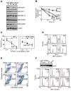
Figure 6. PKA regulation of IAP cytoprotection
A. Modulation of XIAP stability. INS-1 cells expressing GFP, survivin (WT), mitochondrially-targeted survivin (MT-S), or S20A or S20E survivin were exposed to STS, and extracts were analyzed by Western blotting at the indicated time intervals. B. Quantification of XIAP stability. Protein band intensity of the experiment in A was quantified by densitometry. C. Modulation of caspase activity. Recombinant XIAP, WT or S20E survivin was mixed alone (left panel) or in combination (right panel) with Apaf-1, dATP and cytochrome c, and analyzed for DEVDase activity. For combination experiments, a suboptimal concentration of XIAP (0.01 μM) was used. Data are the mean±SEM of three independent experiments. D. DNA content analysis. INS-1 stably transfected with the indicated cDNAs were exposed to STS, and analyzed for DNA content by propidium iodide staining and flow cytometry. The percentage of cells with hypodiploid DNA content is indicated. MT-S, mitochondrially-targeted survivin. E. Multiparametric flow cytometry. INS-1 transfectants stably expressing pcDNA3 (top panels) or S20A or S20E survivin (bottom panels) were treated with STS, and simultaneously analyzed for caspase activity (DEVDase activity) and propidium iodide staining by multiparametric flow cytometry. The percentage of cells in each quadrant is indicated. F. XIAP knockdown. INS-1 cells were transfected with non-targeted dsRNA oligonucleotide (VIII) or increasing concentrations of XIAP-directed siRNA, and analyzed by Western blotting (top panel). *, non specific. Cells stably expressing the indicated constructs were transfected with control (VIII) or XIAP-directed siRNA, exposed to STS, and analyzed for DNA content by propidium iodide staining and flow cytometry (bottom panel). Black line, control siRNA, red line, XIAP siRNA. The percentage of cells with hypodiploid DNA content is indicated.
Figure 7
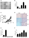
Figure 7. PKA regulated IAP cytoprotection influences tumor growth
A, B. Colony formation assay. Colonies formed in semisolid medium by INS-1 cells stably expressing WT survivin, S20A survivin, mitochondrially-targeted survivin (MT-S) or Bcl-2 were visualized by phase contrast microscopy (A), and scored in individual high-power fields (B). Data are the mean±SEM of four independent experiments. C. Kinetics of tumor growth. INS-1 cells stably expressing the indicated constructs were injected s.c. in the flanks of immunocompromised mice and tumor growth was measured at the indicated time intervals. MT-S, mitochondrially-targeted survivin. Data are the mean±SEM of individual tumor groups (n=8). D. Histology. The various INS-1 tumors in C were analyzed for apoptosis by TUNEL staining, or cell proliferation by Ki67 labeling. Magnification, ×100. E. Apoptotic index. Quantification of TUNEL+ cells in INS-1 tumors. F. Mitotic index. Quantification of Ki67+ cells in INS-1 tumors. For panels E, F, data are the mean±SEM of an average of 800 cells. ***, p<0.0001; ns, not significant.

Similar articles

Cited by

References

    1. Abrams JM. Competition and compensation: coupled to death in development and cancer. Cell. 2002;110:403–406. - PubMed
    1. Altieri DC. Validating survivin as a cancer therapeutic target. Nat Rev Cancer. 2003;3:46–54. - PubMed
    1. Altieri DC. The case for survivin as a regulator of microtubule dynamics and cell-death decisions. Curr Opin Cell Biol. 2006;18:609–615. - PubMed
    1. Amaravadi R, Thompson CB. The survival kinases Akt and Pim as potential pharmacological targets. J Clin Invest. 2005;115:2618–2624. - PMC - PubMed
    1. Amieux PS, McKnight GS. The essential role of RI alpha in the maintenance of regulated PKA activity. Ann N Y Acad Sci. 2002;968:75–95. - PubMed

Publication types

MeSH terms

Substances