. 2007 Jul 6;27(1):79-90.
doi: 10.1016/j.molcel.2007.05.038.

Initiation of RNA decay in Escherichia coli by 5' pyrophosphate removal

Affiliations

Initiation of RNA decay in Escherichia coli by 5' pyrophosphate removal

Helena Celesnik et al. Mol Cell. .

Abstract

The common belief that endonucleolytic cleavage is the initial, rate-determining step of mRNA decay in Escherichia coli fails to explain the influence of 5' termini on the half-lives of primary transcripts. We have re-examined the initial events of RNA degradation in that organism by devising an assay to probe the 5' phosphorylation state of RNA and by employing a self-cleaving hammerhead ribozyme to investigate the degradative consequences of an unphosphorylated 5' end. These studies have identified a previously unrecognized prior step in decay that triggers subsequent internal cleavage by the endonuclease RNase E and thereby governs RNA longevity: the rate-determining conversion of a triphosphorylated to a monophosphorylated 5' terminus. Our findings redefine the role of RNase E in RNA degradation and explain how unpaired 5'-terminal nucleotides can facilitate access to internal cleavage sites within primary transcripts. Moreover, these results reveal a striking parallel between the mechanisms of mRNA decay in prokaryotic and eukaryotic organisms.


Figures

Figure 1
Figure 1. 5’-end-dependent decay of RNA I and 5’ extension variants thereof
(A) RNA I and its derivative RNA I.613. The principal site of RNase E cleavage is marked with an arrow. RNA I+hp and RNA I.613+hp are identical to RNA I and RNA I.613 except for the addition of a 5’-terminal stem-loop (GAUCGCCCACCGGCAGCUGCCGGUGGGCGAUC or AGAGCGGCUUCGGCCGCUCU, respectively) (B) Decay of RNA I, RNA I+hp, RNA I.613, and RNA I.613+hp. At time intervals after inhibiting transcription, total RNA was isolated from E. coli containing plasmid pRNAI, pRNAI+hp, pRNAI.613, or pRNAI.613+hp and analyzed by Northern blotting. tRNACys served as an internal standard. The measured half-lives were 1.8 ± 0.1 min for RNA I, 3.5 ± 0.1 min for RNA I+hp, 0.9 ± 0.1 min for RNA I.613, and 2.8 ± 0.2 min for RNA I.613+hp.
Figure 2
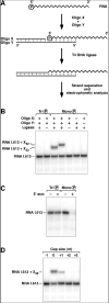
Figure 2. PABLO analysis of RNA synthesized in vitro
(A) Method for analyzing the 5’ phosphorylation state of RNA (PABLO). T4 DNA ligase selectively joins oligo X to 5’-monophosphorylated, but not 5’-triphosphorylated, RNA annealed to the bridging oligo Y, thereby producing an extended DNA-RNA chimera detectable by Northern blotting. (B) Specificity of PABLO. Internally radiolabeled RNA I.613 bearing a 5’ monophosphate or a 5’ triphosphate was prepared by in vitro transcription in the presence or absence of excess AMP. These RNAs were then subjected to PABLO with RNA I.613-specific oligonucleotides, and the effect of changing or omitting various reaction components was determined. The two different oligonucleotides X that were used, X22 (lanes 1, 2, 3, 5, and 7) and X32 (lane 4), were 22 or 32 nucleotides long, respectively. (C) Confirmation of the 5’ phosphorylation state of in vitro transcripts. The 5’ phosphorylation state of the RNA I.613 transcripts examined in (B) was corroborated by digestion with a monophosphate-dependent 5’ exonuclease. (D) Effect of suboptimal end juxtaposition on PABLO ligation yields. The monophosphorylated RNA I.613 sample examined in (B) was subjected to PABLO with a set of bridging Y oligonucleotides that either perfectly juxtaposed the 3’ end of X32 with the 5’ end of the RNA (gap size = 0), left them separated by 1-3 nucleotides (gap size = +1, +2, or +3), or caused them to overlap by one nucleotide (gap size = -1).
Figure 3
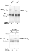
Figure 3. 5’ phosphorylation state of RNA I and RNA I.613 in E. coli
Total RNA extracted from E. coli containing plasmid pRNAI or pRNAI.613 was subjected to PABLO analysis of RNA I or RNA I.613. As a positive control, the 5’ phosphorylation state of the fully monophosphorylated degradation intermediate RNA I-5 was also examined. Oligos X of various lengths were used (XN, where N corresponds to the length in nucleotides). In multiple experiments, the ligation yields were 9 ± 1% and 14 ± 2% for RNA I and RNA I.613, respectively. The radiolabeled probe used to detect RNA I.613 did not detect its 3’ cleavage product (RNA I-5) due to the complementarity of the probe to the 5’-terminal segment of the transcript.
Figure 4
Figure 4. Origin and fate of monophosphorylated RNA I.613 in E. coli
(A) Accumulation of monophosphorylated RNA I.613 when RNase E cleavage is impeded. PABLO was performed to compare the 5’ phosphorylation state of RNA I.613 and RNA I.613-8C, a variant in which the primary RNase E cleavage site of RNA I.613 had been mutated to impede cleavage (ACAGUAUUU → ACCCCCCCC; see Figure 1A). In multiple experiments in E. coli, the ligation yield consistently doubled when RNase E cleavage was inhibited. (B) Phosphorylation state of the 38-nt 5’-terminal decay intermediate produced in E. coli by RNase E cleavage of RNA I.613 (left) or in vitro by RNase E cleavage of fully monophosphorylated, internally radiolabeled RNA I.613 (right). In multiple experiments in E. coli, the PABLO ligation yields were 14 ± 2% and 55 ± 7% for full-length RNA I.613 and its 5’ cleavage product, respectively. (C) Invariant ratio of monophosphorylated to triphosphorylated RNA I.613 following transcription inhibition. (Left) PABLO analysis of RNA I.613 isolated from E. coli at time intervals after inhibiting transcription. (Right) Concentration of monophosphorylated RNA I.613 (ligation product) and triphosphorylated RNA I.613 (unligated) as a function of time after transcription inhibition.
Figure 5
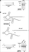
Figure 5. Phosphorylation state of the rpsT P1 transcript and its HO-rpsT counterpart in E. coli
(A) 5’ phosphorylation state of rpsT mRNA. (Left) The E. coli rpsT transcriptional unit. Jagged lines ending in arrowheads, primary transcripts. Gray rectangle, protein-coding region. (Right) PABLO analysis of the chromosomal rpsT P1 transcript in E. coli. In multiple experiments, the ligation yield was 25 ± 2% for the rpsT P1 transcript. (B) Generation of HO-rpsT mRNA by self-cleavage of the hammerhead-containing rpsT.HH transcript. Only the 5’-terminal segment of the transcript is shown. (C) 5’ phosphorylation state of HO-rpsT mRNA. (Left) The rpsT.HH transcriptional unit. (Right) PABLO analysis of HO-rpsT mRNA in E. coli. The plasmid-encoded rpsT P1 transcript and its self-cleaved HO-rpsT P1 counterpart were detected with a radiolabeled probe complementary to a sequence tag inserted into the 3’ UTR.
Figure 6
Figure 6. Stabilization of rpsT mRNA in E. coli by a 5’ hydroxyl
(A) Decay of HO-rpsT mRNA in E. coli. At time intervals after inhibiting transcription, total RNA was isolated from E. coli containing plasmid pRPST1 (top left) or pRPST.HH (bottom left) and analyzed by Northern blotting with a radiolabeled probe complementary to a 3’ UTR sequence tag. Degradation rates were calculated for rpsT P1 mRNA bearing a 5’-terminal hydroxyl (HO-rpsT; half-life = 5.3 ± 0.1 min) or a 5’-terminal triphosphate (TriP-rpsT; half-life = 0.9 ± 0.1 min) by plotting band intensity as a function of time (right). In each case, the triphosphorylated P2 transcript (internal standard) decayed with a half-life of 1.2 ± 0.3 min. (B) Equivalent RNase E cleavage rates in vitro for rpsT RNAs bearing a 5’ triphosphate or a 5’ hydroxyl. Equal amounts of radiolabeled, in vitro synthesized rpsT RNA bearing a 5’ monophosphate, 5’ triphosphate, or 5’ hydroxyl were treated with purified N-RNase E under identical conditions, and samples quenched at time intervals were analyzed by gel electrophoresis and autoradiography.
Figure 7
Figure 7. Mechanism of the 5’-end-dependent pathway for RNA degradation in E. coli
An RNA pyrophosphohydrolase (hatchet) removes the gamma and beta phosphates at the 5’-terminus of a triphosphorylated primary transcript, either simultaneously (as shown) or sequentially. The resulting monophosphorylated decay intermediate is then rapidly cleaved by RNase E (scissors linked to a 5’-sensor domain), an endonuclease with a marked preference for RNA substrates bearing a single phosphate group at the 5’ end.

Similar articles

Cited by

References

    1. Apirion D. Degradation of RNA in Escherichia coli. A hypothesis. Mol Gen Genet. 1973;122:313–322. - PubMed
    1. Arnold TE, Yu J, Belasco JG. mRNA stabilization by the ompA 5’ untranslated region: two protective elements hinder distinct pathways for mRNA degradation. RNA. 1998;4:319–330. - PMC - PubMed
    1. Babitzke P, Kushner SR. The Ams (altered mRNA stability) protein and ribonuclease E are encoded by the same structural gene of Escherichia coli. Proc Natl Acad Sci USA. 1991;88:1–5. - PMC - PubMed
    1. Baker KE, Mackie GA. Ectopic RNase E sites promote bypass of 5’-end-dependent mRNA decay in Escherichia coli. Mol Microbiol. 2003;47:75–88. - PubMed
    1. Baumeister R, Flache P, Melefors Ö, von Gabain A, Hillen W. Lack of a 5’ non-coding region in Tn1721 encoded tetR mRNA is associated with a low efficiency of translation and a short half-life in Escherichia coli. Nucleic Acids Res. 1991;19:4595–4600. - PMC - PubMed

Publication types