. 2007 Jul 6;27(1):149-62.
doi: 10.1016/j.molcel.2007.05.029.

SIRT1 regulates the function of the Nijmegen breakage syndrome protein

Affiliations

SIRT1 regulates the function of the Nijmegen breakage syndrome protein

Zhigang Yuan et al. Mol Cell. .

Abstract

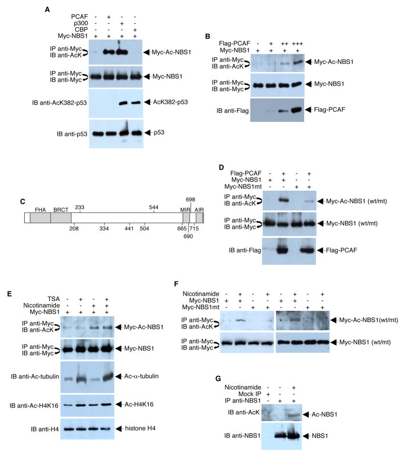
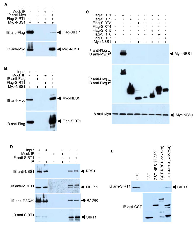
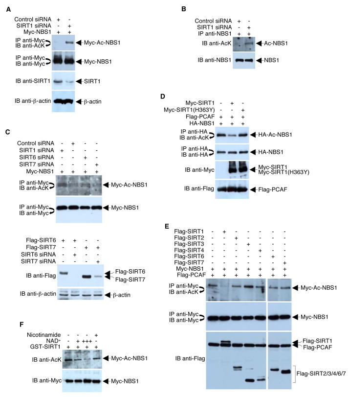
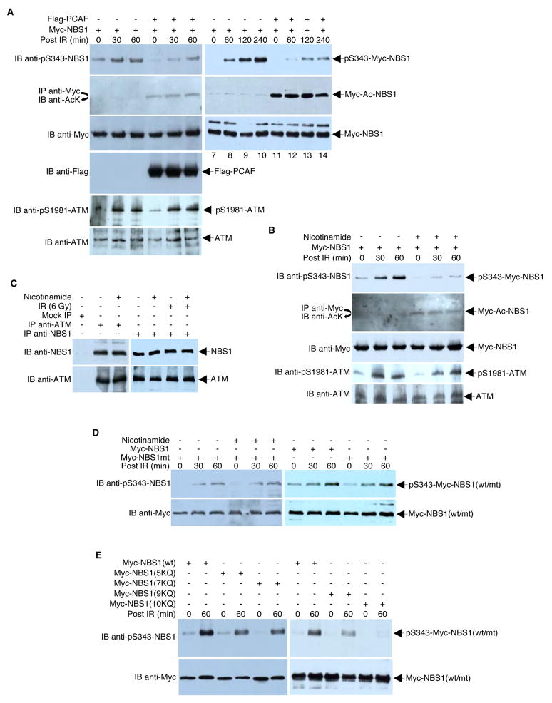
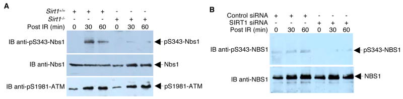
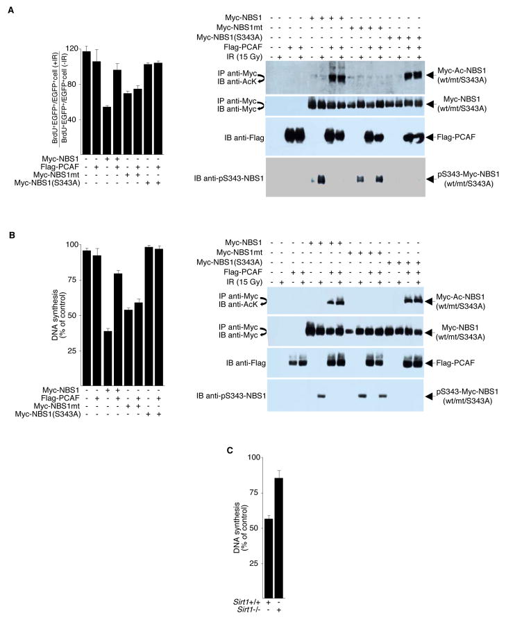
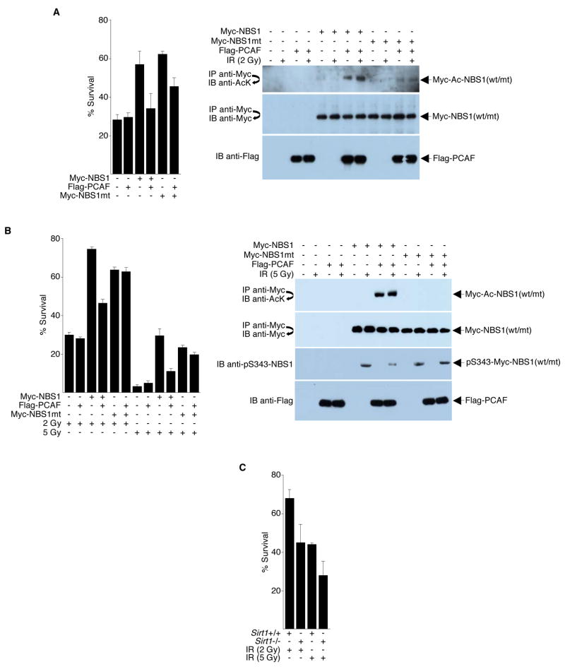
MRE11-RAD50-NBS1 (MRN) is a conserved nuclease complex that exhibits properties of a DNA damage sensor and is critical in regulating cellular responses to DNA double-strand breaks. NBS1, which is mutated in the human genetic disease Nijmegen breakage syndrome, serves as the regulatory subunit of MRN. Phosphorylation of NBS1 by the ATM kinase is necessary for both activation of the S phase checkpoint and for efficient DNA damage repair response. Here, we report that NBS1 is an acetylated protein and that the acetylation level is tightly regulated by the SIRT1 deacetylase. SIRT1 associates with the MRN complex and, importantly, maintains NBS1 in a hypoacetylated state, which is required for ionizing radiation-induced NBS1 Ser343 phosphorylation. Our results demonstrate the presence of crosstalk between two different posttranslational modifications in NBS1 and strongly suggest that deacetylation of NBS1 by SIRT1 plays a key role in the dynamic regulation of the DNA damage response and in the maintenance of genomic stability.


Figures

Figure 1
Figure 1. NBS1 is Acetylated
(A) 293T cells were co-transfected with equal amounts (4 μg) of Myc-NBS1 ± one of the following expression plasmids: Flag-PCAF, HA-p300, or HA-CBP. Cell lysates were immunoprecipitated (IP) under high stringency conditions using anti-Myc antibodies. Immunoprecipitates were subjected to Western blot (IB) analysis using anti-acetyl-lysine (AcK) antibodies. The blot was stripped and re-probed with anti-Myc antibodies to confirm equal immunoprecipitation efficiency and loading. As CBP (but not PCAF) is known to acetylate p53-K382, a Western blot was performed with anti-AcK382-p53 to confirm that the expressed CBP was functional. (B) 293T cells were co-transfected with Myc-NBS1 (4 μg) and Flag-PCAF (0, 0.5, 1, or 4 μg). Cell lysates were subjected to immunoprecipitation/Western blot analysis using the indicated antibodies. Western blot analysis of whole extracts was performed to assess PCAF expression (bottom panel). (C) Myc-NBS1, which was expressed and purified from 293T cells, was digested with trypsin and subjected to ITMS. Positions of the unambiguously-identified acetylated lysine residues are shown. FHA, forkhead-associated domain; BRCT, BRCA1 C-terminal domain; MIR, MRE11-interacting domain; AIR, ATM-interacting domain. (D) 293T cells were co-transfected with Myc-NBS1 (wild-type or lysine to arginine mutant [mt]) and Flag-PCAF (4 μg each), as indicated. Total and acetylated NBS1 levels were analyzed by immunoprecipitation/Western blotting. (E) 293T cells were transfected with plasmids that expressed Myc-NBS1 (4 μg). Twenty-four hours post-transfection, cells were left untreated or treated overnight with TSA (1.3 μM), nicotinamide (20 mM), or TSA plus nicotinamide. Cell lysates were subjected to immunoprecipitation/Western blotting. Whole cell extracts were probed with anti-Ac-tubulin and anti-Ac-H4K16 to confirm that TSA is functional under the treatment condition (lower panels). (F) 293T cells were transfected with plasmids (4 μg each) expressing wild-type or mutant Myc-NBS1 as indicated and then treated with 20 mM nicotinamide overnight. Myc-NBS1 acetylation and protein expression were examined by immunoprecipitation/Western blotting. (G) HeLa cells were treated with nicotinamide (20 mM) or left untreated overnight. Cell lysates were then immunoprecipitated under high stringency conditions with monoclonal anti-NBS1 antibodies. Endogenous acetylated NBS1 was analyzed by Western blotting with a mixture of polyclonal anti-acetyl-lysine antibodies.
Figure 2
Figure 2. SIRT1 Interacts With NBS1
(A, B, C) 293T cells were co-transfected with plasmids (4 μg each) encoding the indicated Myc and Flag fusion proteins. Anti-Myc and anti-Flag immunoprecipitates obtained under low stringency conditions were analyzed by Western blotting with the indicated antibodies. (D) HeLa cells were irradiated (10 Gy) or left untreated. One hour after irradiation, endogenous SIRT1 was immunoprecipitated under low stringency conditions with anti-SIRT1 polyclonal antibodies. Immune complexes were analyzed by Western blotting with anti-NBS1, anti-MRE11, anti-RAD50, or anti-SIRT1 antibodies. For maximum clarity, the image gathered for the left upper middle panel (IB anti-MRE11) was under-exposed compared to the right upper middle panel. (E) GST-NBS1 deletion mutants coupled to Sepharose beads were incubated with whole HeLa cell extracts. After the beads were washed, bound proteins were eluted and analyzed by Western blotting with an anti-SIRT1 antibody. The blot was stripped and re-probed with anti-GST antibodies to confirm equal quantities of GST proteins in each reaction.
Figure 3
Figure 3. SIRT1 Deacetylates NBS1
(A) 293T cells were co-transfected with equal amounts (4 μg) of Myc-NBS1 plasmid and either a plasmid encoding pBS/U6-SIRT1 or control siRNA. Western blots were performed with the indicated antibodies to assess the levels of SIRT1, β-actin, total Myc-NBS1, and acetylated Myc-NBS1. (B) 293T cells were infected with either adenovirus that expresses control siRNA or adenovirus that expresses SIRT1 siRNA. Western blots were performed with the indicated antibodies to assess the acetylation of endogenous NBS1 and NBS1 immunoprecipitation efficiency. (C) Upper panels, 293T cells were co-transfected with equal amounts (4 μg) of Myc-NBS1 plasmid and either a plasmid encoding SIRT1 siRNA, SIRT6 siRNA, SIRT7 siRNA, or control siRNA. Western blots were performed with the indicated antibodies to assess the levels of total Myc-NBS1 and acetylated Myc-NBS1. Bottom panels, similar experiments were performed with over-expression of Flag-SIRT6 and Flag-SIRT7 to show that SIRT6 and SIRT7 siRNAs are functional. D) 293T cells were co-transfected with plasmids that express HA-NBS1, Flag-PCAF, and either a wild-type or a catalytically-defective Myc-SIRT1. Acetylation of HA-NBS1 and all protein levels were determined with direct Western blotting or immunoprecipitations followed by Western blotting using the indicated antibodies. (E) 293T cells were co-transfected with plasmids that express Myc-NBS1, Flag-PCAF, and different Flag-tagged SIRTs. Acetylation of Myc-NBS1 and all protein levels were determined with direct Western blotting or immunoprecipitations followed by Western blotting using the indicated antibodies. (F) 293T cells were transfected with Myc-NBS1 and Flag-PCAF. Anti-Myc immunoprecipitates were incubated with recombinant GST-SIRT1 in the presence or absence of NAD+ (+, 1mM; +++, 10 mM) and nicotinamide (10 mM) at 30°C for 1 h. Western blot analysis of acetylated Myc-NBS1 was then performed using anti-acetyl-lysine (AcK) antibodies.
Figure 4
Figure 4. NBS1 Acetylation Status Affects IR-induced NBS1 Phosphorylation
(A) The NBS cell line GM07166 was transfected with equal amounts (4 μg) of Myc-NBS1 expression plasmid and Flag-PCAF expression plasmid or empty vector. Twenty-four hours post-transfection, cells were irradiated (6 Gy) or left untreated. Cell lysates were subjected to Western blot analysis or to high stringency immunoprecipitation/Western blot analysis, as indicated. (B) NBS cells transfected with Myc-NBS1 expression plasmid (4 μg) were treated overnight with 20 mM of nicotinamide or left untreated. The cells were then irradiated (6 Gy) or left untreated. Cell lysates were prepared after the indicated time and subjected to direct Western blot analysis or high stringency immunoprecipitation/Western blot analysis using the indicated antibodies. (C) To examine the effect of nicotinamide on ATM-NBS1 interactions, ATM and NBS1 were immunoprecipitated with anti-ATM antibodies and anti-NBS1 antibodies, respectively, from extracts prepared from HeLa cells treated ± nicotinamide. NBS1 and ATM were identified in the immunoprecipitates by Western blotting with anti-NBS1 and anti-ATM, respectively. (D) NBS cells were transfected with plasmids encoding either wild-type or acetylation-defective Myc-NBS1 (4 μg). Parallel cultures were then irradiated or left unirradiated in the presence or absence of nicotinamide. Lysates were analyzed by Western blots with the indicated antibodies. (E) NBS cells were transfected with plasmids encoding either wild-type Myc-NBS1 or acetylation-mimic Myc-NBS1 mutants (4 μg). Parallel cultures were then irradiated or left unirradiated and lysates were analyzed by Western blots with the indicated antibodies.
Figure 5
Figure 5. Presence of SIRT1 affects NBS1 Ser343 phosphorylation
(A) Murine Sirt1+/+ and Sirt1−/− fibroblasts were irradiated (6 Gy) or left unirradiated. At different time points after IR, cell lysates were collected and analyzed by Western blotting with anti-pS343-mNbs1 antibodies. The blot was sequentially stripped and re-probed with anti-mNbs1 and pS1981-ATM antibodies to confirm equal mNbs1 protein levels and intact ATM activation. (B) 293T cells infected with adenoviruses expressing SIRT1 siRNA or control viruses were irradiated (6 Gy) or left unirradiated. At different time points after IR, cell lysates were collected and analyzed by Western blotting with anti-pS343-NBS1 antibodies. The blot was stripped and re-probed with anti-NBS1.
Figure 6
Figure 6. NBS1 Acetylation Affects Radioresistant DNA synthesis (RDS)
(A) NBS cells were transfected with plasmids encoding the indicated proteins. Twenty-four hours after transfection, cells were irradiated (15 Gy) or left unirradiated. Left panel, RDS was determined by comparing the ratio of BrdU+/EGFP+ cells to EGFP+ cells. The bar represents the ratio in the IR-treated cells divided by the ratio in the untreated cells. Right panels, to assess immunoprecipitation efficiency, protein expression, and NBS1 acetylation/phosphorylation, cell lysates were subjected to Western blot analysis or to high stringency immunoprecipitation/Western blot analysis, as indicated. Representative blots are shown. (B) Left panel, stably transfected NBS cells (GM 07166) were treated with 15 Gy of IR or left untreated, cells were radiolabeled with thymidine for RDS analysis. Right panels, representative blots to monitor immunoprecipitation efficiency, protein expression, and NBS1 acetylation/phosphorylation. (C) 2 × 10⁴ Sirt1+/+ or Sirt1−/− MEFs were plated on 35 mm dishes for 48 h. RDS assays were performed using the radio-labeling method to determine inhibition of DNA synthesis after 15 Gy of IR. Error bars denote the standard deviation (SD). The data are expressed as mean ± SD from three separate experiments.
Figure 7
Figure 7. NBS1 Acetylation Affects Cell Survival
(A) Left panel, NBS cells transfected with plasmids (4 μg each) encoding the indicated proteins were irradiated (2 Gy) or left unirradiated. The surviving colonies were counted two weeks later. Right panels, direct Western blots and high stringency immunoprecipitation/Western blot analyses were performed to determine NBS1 acetylation, immunoprecipitation efficiency, and protein expression levels. Representative blots are shown. (B) Left panel, stably transfected NBS cells (GM 07166) were treated with 2 or 5 Gy of IR or left untreated and surviving colonies were counted two weeks later. Right panels, direct Western blots and high stringency immunoprecipitation/Western blot analyses were performed to determine NBS1 acetylation/phosphorylation, immunoprecipitation efficiency, and protein expression levels. Although similar results were obtained from 2 Gy and 5 Gy IR treatments, representative blots from 5 Gy treatments only are shown for simplicity. (C) Sirt1+/+ and Sirt1−/− MEFs were irradiated with 2 or 5 Gy of IR or left unirradiated. The surviving colonies were counted two weeks later. Fractional cell survival is the fraction of colonies surviving irradiation divided by the total number of colonies in the unirradiated parallel culture. Error bars denote the standard deviation (SD). The data are expressed as mean ± SD from three separate experiments.

Similar articles

Cited by

References

    1. Bakkenist CJ, Kastan MB. DNA damage activates ATM through intermolecular autophosphorylation and dimer dissociation. Nature. 2003;421:499–506. - PubMed
    1. Blander G, Guarente L. The Sir2 family of protein deacetylases. Annu Rev Biochem. 2004;73:417–435. - PubMed
    1. Blander G, Olejnik J, Krzymanska-Olejnik E, McDonagh T, Haigis M, Yaffe MB, Guarente L. SIRT1 shows no substrate specificity in vitro. J Biol Chem. 2005;280:9780–9785. - PubMed
    1. Boisvert FM, Dery U, Masson JY, Richard S. Arginine methylation of MRE11 by PRMT1 is required for DNA damage checkpoint control. Genes Dev. 2005;19:671–676. - PMC - PubMed
    1. Brand M, Moggs JG, Oulad-Abdelghani M, Lejeune F, Dilworth FJ, Stevenin J, Almouzni G, Tora L. UV-damaged DNA-binding protein in the TFTC complex links DNA damage recognition to nucleosome acetylation. EMBO J. 2001;20:3187–3196. - PMC - PubMed

Publication types