

N-acetyl aspartate in autism spectrum disorders: regional effects and relationship to fMRI activation
- PMID: 17612510
- PMCID: PMC3477551
- DOI: 10.1016/j.brainres.2007.04.081
N-acetyl aspartate in autism spectrum disorders: regional effects and relationship to fMRI activation
Abstract
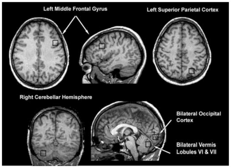
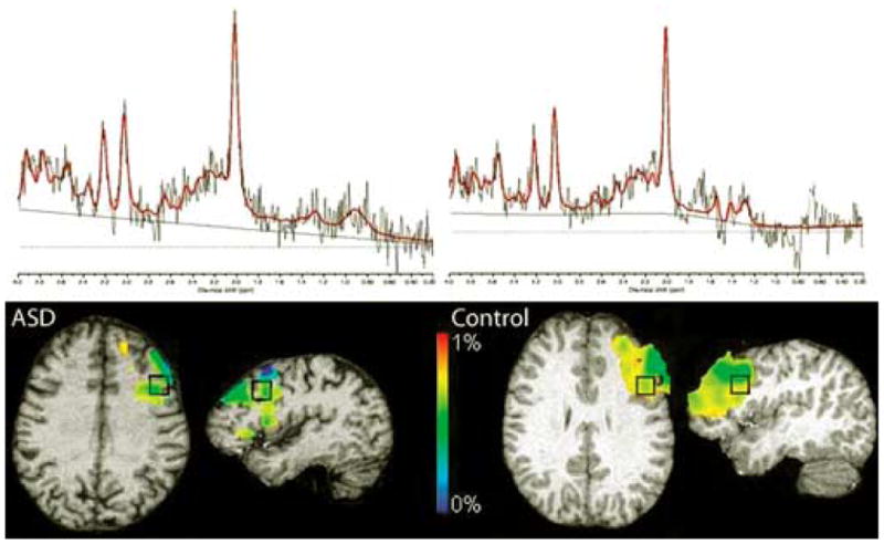
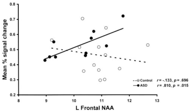
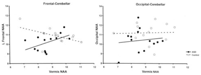
Rapid progress in our understanding of macrostructural abnormalities in autism spectrum disorders (ASD) has occurred in recent years. However, the relationship between the integrity of neural tissue and neural function has not been previously investigated. Single-voxel proton magnetic resonance spectroscopy and functional magnetic resonance imaging of an executive functioning task was obtained in 13 high functioning adolescents and adults with ASD and 13 age-matched controls. The ASD group showed significant reductions in N-acetyl aspartate (NAA) in all brain regions combined and a specific reduction in left frontal cortex compared to controls. Regression analyses revealed a significant group interaction effect between frontal and cerebellar NAA. In addition, a significant positive semi-partial correlation between left frontal lobe NAA and frontal lobe functional activation was found in the ASD group. These findings suggest that widespread neuronal dysfunction is present in high functioning individuals with ASD. Hypothesized developmental links between frontal and cerebellar vermis neural abnormalities were supported, in that impaired neuronal functioning in the vermis was associated with impaired neuronal functioning in the frontal lobes in the ASD group. Furthermore, this study provided the first direct evidence of the relationship between abnormal functional activation in prefrontal cortex and neuronal dysfunction in ASD.
Figures

Similar articles
-
Altered chemical metabolites in the amygdala-hippocampus region contribute to autistic symptoms of autism spectrum disorders.Biol Psychiatry. 2007 Nov 1;62(9):1030-7. doi: 10.1016/j.biopsych.2007.05.015. Epub 2007 Jul 12. Biol Psychiatry. 2007. PMID: 17631869
-
Proton magnetic resonance spectroscopy of the frontal lobe and cerebellar vermis in children with a mood disorder and a familial risk for bipolar disorders.J Child Adolesc Psychopharmacol. 2003 Winter;13(4):545-55. doi: 10.1089/104454603322724931. J Child Adolesc Psychopharmacol. 2003. PMID: 14977467 Clinical Trial.
-
Biochemistry of the cingulate cortex in autism: An MR spectroscopy study.Autism Res. 2016 Jun;9(6):643-57. doi: 10.1002/aur.1562. Epub 2015 Nov 3. Autism Res. 2016. PMID: 26526126 Free PMC article.
-
Proton magnetic resonance spectroscopy as a probe into the pathophysiology of autism spectrum disorders (ASD): a review.Autism Res. 2013 Apr;6(2):119-33. doi: 10.1002/aur.1273. Epub 2013 Feb 21. Autism Res. 2013. PMID: 23436782 Review.
-
Neuroimaging in autism spectrum disorders: 1H-MRS and NIRS study.J Med Invest. 2015;62(1-2):29-36. doi: 10.2152/jmi.62.29. J Med Invest. 2015. PMID: 25817280 Review.
Cited by
-
Brain Metabolite Changes After Anodal Transcranial Direct Current Stimulation in Autism Spectrum Disorder.Front Mol Neurosci. 2020 Jun 4;13:70. doi: 10.3389/fnmol.2020.00070. eCollection 2020. Front Mol Neurosci. 2020. PMID: 32581703 Free PMC article.
-
Elevated glutamatergic compounds in pregenual anterior cingulate in pediatric autism spectrum disorder demonstrated by 1H MRS and 1H MRSI.PLoS One. 2012;7(7):e38786. doi: 10.1371/journal.pone.0038786. Epub 2012 Jul 27. PLoS One. 2012. PMID: 22848344 Free PMC article. Clinical Trial.
-
Age-related change in brain metabolite abnormalities in autism: a meta-analysis of proton magnetic resonance spectroscopy studies.Transl Psychiatry. 2012 Jan 17;2(1):e69. doi: 10.1038/tp.2011.65. Transl Psychiatry. 2012. PMID: 22832731 Free PMC article.
-
A Magnetic Resonance Spectroscopy Study of Superior Visual Search Abilities in Children with Autism Spectrum Disorder.Autism Res. 2020 Apr;13(4):550-562. doi: 10.1002/aur.2258. Epub 2020 Jan 7. Autism Res. 2020. PMID: 31909886 Free PMC article.
-
Fragile X Syndrome FMRP Co-localizes with Regulatory Targets PSD-95, GABA Receptors, CaMKIIα, and mGluR5 at Fiber Cell Membranes in the Eye Lens.Neurochem Res. 2015 Nov;40(11):2167-76. doi: 10.1007/s11064-015-1702-2. Epub 2015 Aug 23. Neurochem Res. 2015. PMID: 26298628
References
-
- Akshoomoff NA, et al. Contribution of the cerebellum to neuropsychological functioning: evidence from a case of cerebellar degenerative disorder. Neuropsychologia. 1992;30:315–28. - PubMed
-
- Allen G, Courchesne E. Differential effects of developmental cerebellar abnormality on cognitive and motor functions in the cerebellum: an fMRI study of autism. Am J Psychiatry. 2003;160:262–73. - PubMed
-
- Allen G, et al. Cerebellar function in autism: functional magnetic resonance image activation during a simple motor task. Biol Psychiatry. 2004;56:269–78. - PubMed
-
- Aylward EH, et al. Effects of age on brain volume and head circumference in autism. Neurology. 2002;59:175–83. - PubMed
Publication types
MeSH terms
Substances
Grants and funding
LinkOut - more resources
Full Text Sources
Other Literature Sources
Medical
