

Distinct cardiodynamic and molecular characteristics during early and late stages of sepsis-induced myocardial dysfunction
- PMID: 17612571
- PMCID: PMC1986677
- DOI: 10.1016/j.lfs.2007.05.021
Distinct cardiodynamic and molecular characteristics during early and late stages of sepsis-induced myocardial dysfunction
Abstract
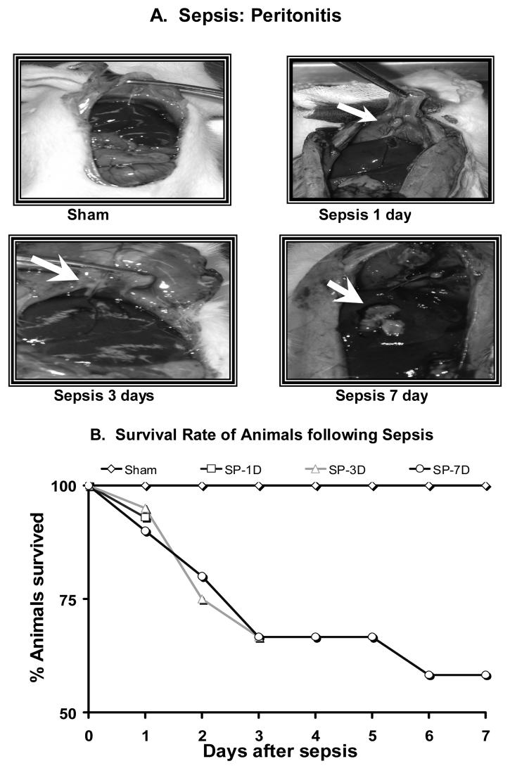
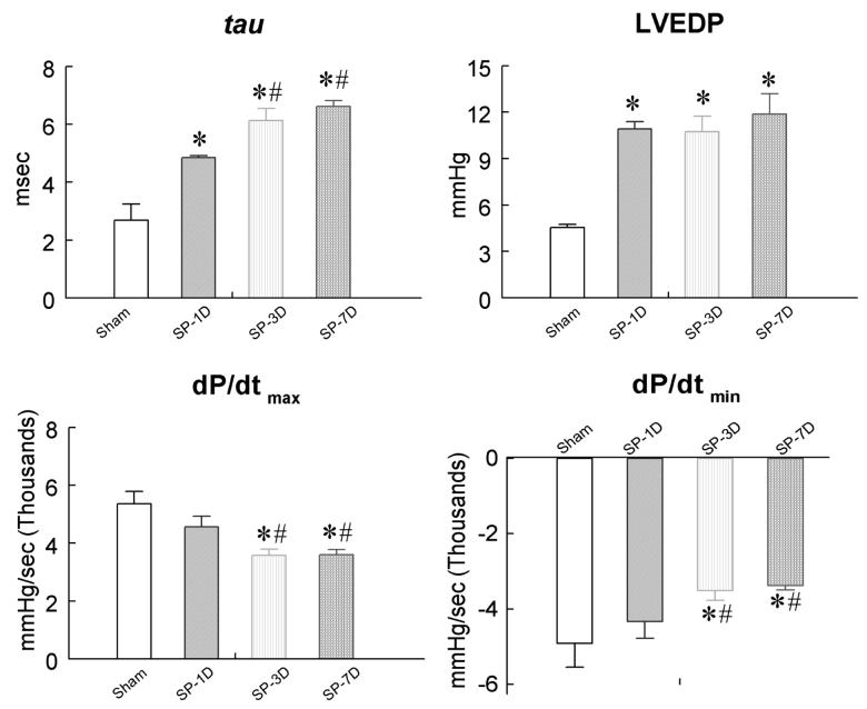
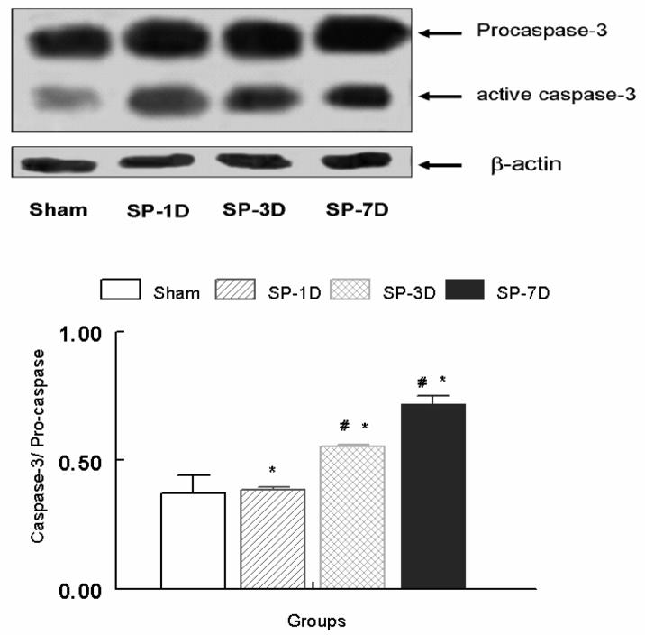
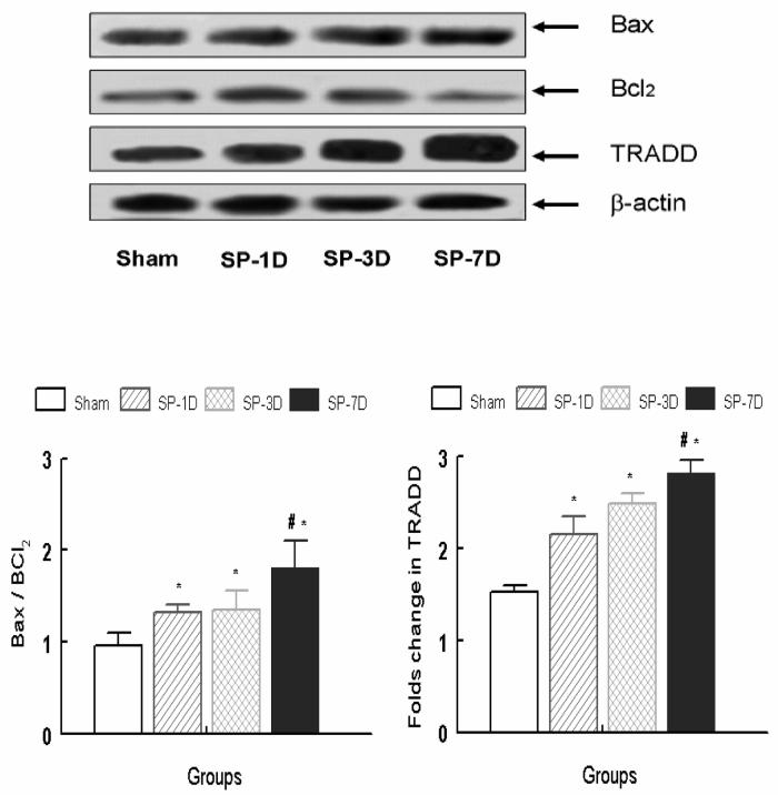
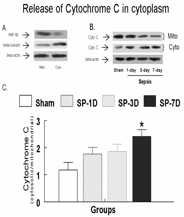
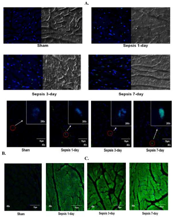
We hypothesized that progressive decline in myocardial performance would correlate with upregulation of markers for apoptotic mechanisms following increased duration of polymicrobial sepsis in the rat. Male Sprague-Dawley rats (350-400 g) were randomized into sham, 1-, 3- and 7-day sepsis groups. Each septic rat received 200 mg/kg cecal inoculum intraperitoneally (i.p). The post-mortem analysis showed a severely inflamed peritoneum with the presence of pus in all septic animals that was directly proportional to the duration of sepsis. We observed 10, 33 and 42% mortality in the 1-, 3- and 7-day sepsis groups, respectively. Septic animals at 3 and 7 days exhibited an increased wet lung/total body weight and heart weight/total body weight. A significant increase in total cardiac troponin I (cTnI) and C Reactive Protein (CRP) and endothelin-1 (ET-1) was also observed with an increased duration of sepsis. Myocardial ET-1 concentration in the 7-day post-sepsis group was significantly elevated compared to the sham and 1-day post-sepsis groups. Sepsis also produced a significant decrease in the mean arterial pressure in the 7-day post-sepsis group and tachycardia in the 1-, 3-, and 7-day post-sepsis groups compared to the sham group. A significant prolongation of the left ventricular isovolumic relaxation rate constant, tau, and left ventricular end-diastolic pressure in the 1-, 3- and 7-day post-sepsis groups compared to the sham group was observed. In addition, a significant decrease in the rates of left ventricular relaxation (-dP/dt) and contraction (+dP/dt) in the 3- and 7-day post-sepsis groups compared to the sham and 1-day post-sepsis group was observed. Sepsis produced a significant upregulation in the expression of myocardial TRADD, cytosolic active caspase-3, the Bax/Bcl(2) ratio, and the mitochondrial release of cytochrome C in the 3- and 7-day post-sepsis groups. We observed a progressive increase in the number of TUNEL positive nuclei, cytosolic caspase-3 activation and co-localization of PARP in the nuclei at 1, 3 and 7 days post-sepsis. These data suggest that the progression of sepsis from 1 day to 3-7 days produce distinct cardiodynamic characteristics with a more profound effect during later stages. The sepsis-induced decline in myocardial performance correlates with the induction of myocardial apoptosis.
Figures
Similar articles
-
Alpha-1 Adrenergic Receptor Agonist Phenylephrine Inhibits Sepsis-Induced Cardiomyocyte Apoptosis and Cardiac Dysfunction via Activating ERK1/2 Signal Pathway.Shock. 2019 Jul;52(1):122-133. doi: 10.1097/SHK.0000000000001205. Shock. 2019. PMID: 29889817
-
Sepsis-induced myocardial dysfunction.Shock. 2007 Sep;28(3):265-9. doi: 10.1097/01.shk.0000235090.30550.fb. Shock. 2007. PMID: 17529910 Review.
-
Modulation of myocardial mitochondrial mechanisms during severe polymicrobial sepsis in the rat.PLoS One. 2011;6(6):e21285. doi: 10.1371/journal.pone.0021285. Epub 2011 Jun 21. PLoS One. 2011. PMID: 21712982 Free PMC article.
-
Bigendothelin-1 (1-21) fragment during early sepsis modulates tau, p38-MAPK phosphorylation and nitric oxide synthase activation.Mol Cell Biochem. 2005 Mar;271(1-2):225-37. doi: 10.1007/s11010-005-6416-3. Mol Cell Biochem. 2005. PMID: 15881674
-
Chronic peritoneal sepsis: myocardial dysfunction, endothelin and signaling mechanisms.Front Biosci. 2005 Sep 1;10:3183-205. doi: 10.2741/1774. Front Biosci. 2005. PMID: 15970572 Review.
Cited by
-
Upregulation of myocardial syntaxin1A is associated with an early stage of polymicrobial sepsis.Mol Cell Biochem. 2009 Mar;323(1-2):61-8. doi: 10.1007/s11010-008-9964-5. Epub 2008 Nov 28. Mol Cell Biochem. 2009. PMID: 19039652
-
Contractile response of norepinephrine is modulated by caspase-3 in adult rat ventricular myocytes isolated from septic rat heart.Pharmacol Res. 2009 Oct;60(4):303-13. doi: 10.1016/j.phrs.2009.04.009. Epub 2009 Apr 24. Pharmacol Res. 2009. PMID: 19394424 Free PMC article.
-
Clemastine protects against sepsis-induced myocardial injury in vivo and in vitro.Bioengineered. 2022 Mar;13(3):7134-7146. doi: 10.1080/21655979.2022.2047256. Bioengineered. 2022. PMID: 35274595 Free PMC article.
-
Caspase-3 knock-down reverses contractile dysfunction induced by sepsis in adult rat ventricular myocytes.Br J Pharmacol. 2010 May;160(1):93-100. doi: 10.1111/j.1476-5381.2010.00686.x. Epub 2010 Mar 19. Br J Pharmacol. 2010. PMID: 20331606 Free PMC article.
-
Signaling mechanism of poly(ADP-ribose) polymerase-1 (PARP-1) in inflammatory diseases.Am J Pathol. 2011 Mar;178(3):946-55. doi: 10.1016/j.ajpath.2010.12.004. Am J Pathol. 2011. PMID: 21356345 Free PMC article. Review.
References
-
- Ammann P, Fehr T, Minder EI, Gunter C, Bertel O. Elevation of troponin I in sepsis and septic shock. Intensive Care Medicine. 2001;27:965–969. - PubMed
-
- Arlati S, Brenna S, Prencipe L, Marocchi A, Casella GP, Lanzani M, Gandini C. Myocardial necrosis in ICU patients with acute non-cardiac disease: a prospective study. Intensive Care Medicine. 2000;26:31–37. - PubMed
-
- Behr TM, Nerurkar SS, Nelson AH, Coatney RW, Woods TN, Sulpizio A, Chandra S, Brooks DP, Kumar S, Lee JC, Ohlstein EH, Angermann CE, Adams JL, Sisko J, Sackner-Bernstein JD, Willette RN. Hypertensive end-organ damage and premature mortality are p38 mitogen-activated protein kinase-dependent in a rat model of cardiac hypertrophy and dysfunction. Circulation. 2001;104:1292–1298. - PubMed
-
- Bertinchant JP, Larue C, Pernel I, Ledermann B, Fabbro-Peray P, Beck L, Calzolari C, Trinquier S, Nigond J, Pau B. Release kinetics of serum cardiac troponin I in ischemic myocardial injury. Clinical Biochemistry. 1996;29:587–594. - PubMed
-
- Brahmbhatt S, Gupta A, Sharma AC. Bigendothelin-1 (1-21) fragment during Early Sepsis Modulates tau, p38-MAPK phosphorylation and Nitric Oxide Synthase Activation. Molecular and Cellular Biochemistry. 2005;271:225–237. - PubMed
Publication types
MeSH terms
Substances
Grants and funding
LinkOut - more resources
Full Text Sources
Medical
Research Materials
Miscellaneous
