

doi: 10.1007/BF03085955.
Clinical usefulness of SonoVue contrast echocardiography: the Thoraxcentre experience
Affiliations
- PMID: 17612661
- PMCID: PMC1847751
- DOI: 10.1007/BF03085955
Item in Clipboard
Clinical usefulness of SonoVue contrast echocardiography: the Thoraxcentre experience
Neth Heart J.
2007.
Abstract
Although other imaging techniques, such as magnetic resonance imaging and computer tomography, are becoming more and more important in cardiology, two-dimensional echocardiography is still the most used technique in clinical cardiology. Quantification of left ventricular function and dimensions is important because therapeutic strategies, for example implanting an ICD after myocardial infarction, are based on ejection fraction measurements. Because of the sometimes low quality of echocardiographic images we started to use an ultrasound contrast agent and in this article we describe our experiences with SonoVue, a second-generation contrast agent, over a threeyear period in the Thoraxcentre. (Neth Heart J 2007;15:55-60.).
Figures

SonoVue file including syringe and needle.

Upper panel shows myocardial reflection and lower panel microbubble reflection in power modulation mode (also see text for explanation). From: Mor-Avi V, et al. Circulation 2001;104:352-7.
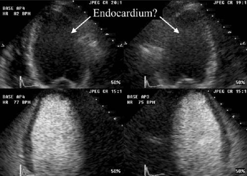
Visualisation of endocardial border in four- and three-chamber images without (upper images) and with (lower images) contrast.

Intertrabecular recesses filling with contrast (*) and apical mobile thrombus in a patient with noncompaction cardiomyopathy.

Endocardial border detection of right ventricle without (left) and with contrast (right).

Patient with echinococcus cyst; contrast image shows no connection with left ventricle.
Similar articles
-
Italian Society of Cardiovascular Echography (SIEC) Consensus Conference on the state of the art of contrast echocardiography.Ital Heart J. 2004 Apr;5(4):309-34. Ital Heart J. 2004. PMID: 15185894 Review.
-
[Estimation of left ventricular volumes and ejection fraction with acoustic quantification in myocardial infarction. Comparison with echocardiographic, angiographic and scintigraphic data].Arch Mal Coeur Vaiss. 1996 Jul;89(7):843-9. Arch Mal Coeur Vaiss. 1996. PMID: 8869245 French.
-
European Association of Cardiovascular Imaging/Cardiovascular Imaging Department of the Brazilian Society of Cardiology recommendations for the use of cardiac imaging to assess and follow patients after heart transplantation.Eur Heart J Cardiovasc Imaging. 2015 Sep;16(9):919-48. doi: 10.1093/ehjci/jev139. Epub 2015 Jul 2. Eur Heart J Cardiovasc Imaging. 2015. PMID: 26139361 Review.
-
Comparison of contrast agent-enhanced versus non-contrast agent-enhanced real-time three-dimensional echocardiography for analysis of left ventricular systolic function.Am J Cardiol. 2007 Nov 1;100(9):1485-9. doi: 10.1016/j.amjcard.2007.06.042. Epub 2007 Aug 24. Am J Cardiol. 2007. PMID: 17950813
-
Usefulness of Speckle-Tracking Imaging for Right Ventricular Assessment after Acute Myocardial Infarction: A Magnetic Resonance Imaging/Echocardiographic Comparison within the Relation between Aldosterone and Cardiac Remodeling after Myocardial Infarction Study.J Am Soc Echocardiogr. 2015 Jul;28(7):818-27.e4. doi: 10.1016/j.echo.2015.02.019. Epub 2015 Mar 31. J Am Soc Echocardiogr. 2015. PMID: 25840640 Clinical Trial.
Cited by
-
Echo contrast medium: How the use of contrast echocardiography (ultrasound contrast agents) can improve patient care.World J Methodol. 2025 Sep 20;15(3):100490. doi: 10.5662/wjm.v15.i3.100490. eCollection 2025 Sep 20. World J Methodol. 2025. PMID: 40881222 Review.
-
Ultrasound imaging beyond the vasculature with new generation contrast agents.Wiley Interdiscip Rev Nanomed Nanobiotechnol. 2015 Jul-Aug;7(4):593-608. doi: 10.1002/wnan.1326. Epub 2015 Jan 8. Wiley Interdiscip Rev Nanomed Nanobiotechnol. 2015. PMID: 25580914 Free PMC article. Review.
-
Ultrafast optical and passive acoustic mapping characterization of nanoscale cavitation nuclei based on gas vesicle proteins.AIP Adv. 2025 Feb 7;15(2):025016. doi: 10.1063/5.0239607. eCollection 2025 Feb. AIP Adv. 2025. PMID: 39944082 Free PMC article.
-
Adverse reactions after the use of SonoVue contrast agent: Characteristics and nursing care experience.Medicine (Baltimore). 2019 Nov;98(44):e17745. doi: 10.1097/MD.0000000000017745. Medicine (Baltimore). 2019. PMID: 31689827 Free PMC article.
References
-
- Hundley WG, Kizilbash AM, Afridi I, Franco F, Peshock RM, Grayburn PA. Administration of an intravenous perfluorocarbon contrast agent improves echocardiographic determination of left ventricular volumes and ejection fraction: comparison with cine magnetic resonance imaging. J Am Coll Cardiol 1998;32:1426- 32. - PubMed
-
- Kasprzak JD, Paelinck B, Ten Cate FJ, et al. Comparison of native and contrast-enhanced harmonic echocardiography for visualization of left ventricular endocardial border. Am J Cardiol 1999;83:211-7. - PubMed
-
- Sieswerda GT, Kamp O, Visser CA. The use of contrast agents in echocardiography. Part I: History, Principles and Developments in Agents and Ultrasound Technology. Neth Heart J 1998;5:583-8.
-
- Sieswerda GT, Kamp O, Visser CA. The use of contrast agents in echocardiography. Part II: clinical indications and experimental applications. Neth Heart J 1998;5:648-57.
-
- Schneider M. SonoVue, a new ultrasound contrast agent. Eur Radiol 1999;9(Suppl 3):S347-8. - PubMed
LinkOut - more resources
Full Text Sources