Primary monophasic mediastinal, cardiac and pericardial synovial sarcoma: a young man in distress
- PMID: 17612689
- PMCID: PMC1896143
- DOI: 10.1007/BF03085986
Primary monophasic mediastinal, cardiac and pericardial synovial sarcoma: a young man in distress
Abstract
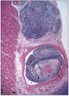
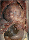
A 19-year-old male was admitted because of exertional dyspnoea. The imaging studies revealed epicardial, pericardial and mediastinal masses. The tumours could not be resected through a minor thoracotomy, only biopsies could be taken. Analyses led to the final diagnosis of a monophasic synovial sarcoma. The patient preferred a conservative and palliative approach. Three months later he died at home. Autopsy demonstrated dramatic extension of the tumour masses. We conclude this report with a discussion on primary cardiac tumours. (Neth Heart J 2007;15:226-8.).
Figures


References
-
- Burke AP, Cowan D, Virmani R. Primary sarcomas of the heart. Cancer 1992;69:387-95. - PubMed
-
- Donsbeck AV, Ranchere D, Coindre JM, Le Gall F, Cordier JF, Loire R. Primary cardiac sarcomas: an immunohistochemical and grading study with long-term follow-up of 24 cases. Histopathology 1999;34:295-304. - PubMed
-
- Nicholson AG, Rigby M, Lincoln C, Meller S, Fisher C. Synovial sarcoma of the heart. Histopathology 1997;30:349-52. - PubMed
-
- Weiss SW, Goldblum JR. Malignant soft tissue tumors of uncertain type. In: Enzinger and Weiss’s soft tissue tumors, 4th ed. St. Louis, MO; Mosby, 2001:1483-509.
-
- McGilbray TT, Schulz TK. Clinical picture: Primary cardiac synovial sarcoma. Lancet Oncol 2003;4:283. - PubMed
LinkOut - more resources
Full Text Sources
