

Alteration of iron regulatory proteins (IRP1 and IRP2) and ferritin in the brains of scrapie-infected mice
- PMID: 17614197
- PMCID: PMC2365884
- DOI: 10.1016/j.neulet.2007.05.061
Alteration of iron regulatory proteins (IRP1 and IRP2) and ferritin in the brains of scrapie-infected mice
Abstract
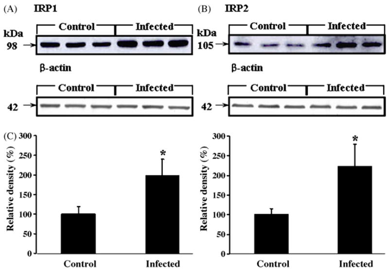
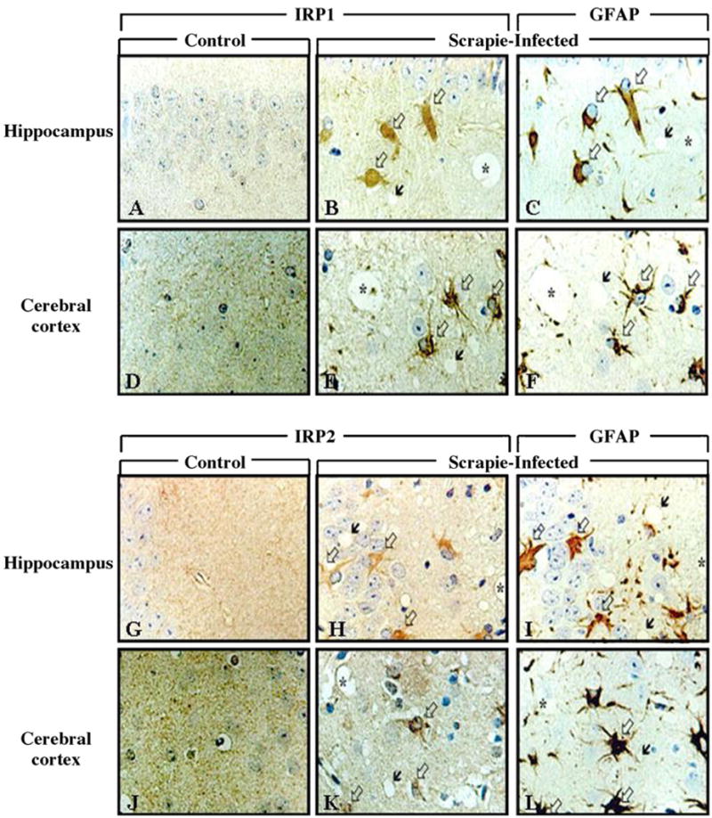
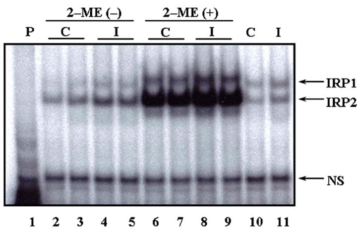
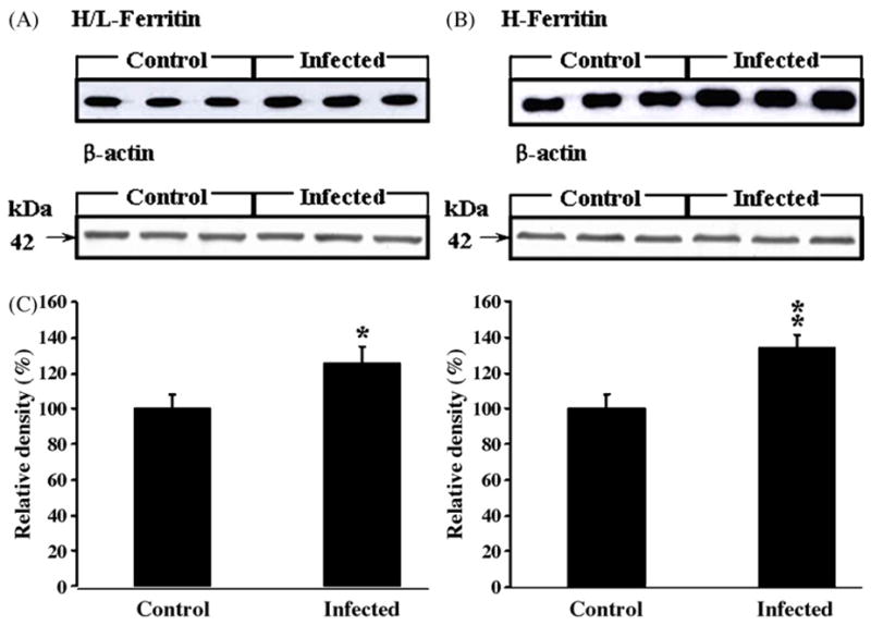
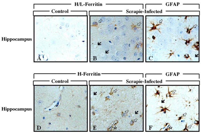
Considerable evidence suggests that oxidative stress may be involved in the pathogenesis of Transmissible Spongiform Encephalopathies (TSEs). To investigate the involvement of iron metabolism in TSEs, we examined the expression levels of iron regulatory proteins (IRPs), ferritins, and binding activities of IRPs to iron-responsive element (IRE) in scrapie-infected mice. We found that the IRPs-IRE-binding activities and ferritins were increased in the astrocytes of hippocampus and cerebral cortex in the brains of scrapie-infected mice. These results suggest that alteration of iron metabolism contributes to development of neurodegeneration and that some protective mechanisms against iron-induced oxidative damage may occur during the pathogenesis of TSEs.
Figures
Similar articles
-
Changed iron regulation in scrapie-infected neuroblastoma cells.Brain Res Mol Brain Res. 2005 Feb 18;133(2):266-73. doi: 10.1016/j.molbrainres.2004.10.018. Brain Res Mol Brain Res. 2005. PMID: 15710243
-
Excess capacity of the iron regulatory protein system.J Biol Chem. 2007 Aug 24;282(34):24650-9. doi: 10.1074/jbc.M703167200. Epub 2007 Jun 28. J Biol Chem. 2007. PMID: 17604281
-
Mammalian tissue oxygen levels modulate iron-regulatory protein activities in vivo.Science. 2004 Dec 17;306(5704):2087-90. doi: 10.1126/science.1103786. Science. 2004. PMID: 15604406
-
Mammalian iron metabolism and its control by iron regulatory proteins.Biochim Biophys Acta. 2012 Sep;1823(9):1468-83. doi: 10.1016/j.bbamcr.2012.05.010. Epub 2012 May 17. Biochim Biophys Acta. 2012. PMID: 22610083 Free PMC article. Review.
-
Role of nitric oxide in cellular iron metabolism.Biometals. 2003 Mar;16(1):125-35. doi: 10.1023/a:1020788603046. Biometals. 2003. PMID: 12572672 Review.
Cited by
-
Key players in the regulation of iron homeostasis at the host-pathogen interface.Front Immunol. 2023 Oct 24;14:1279826. doi: 10.3389/fimmu.2023.1279826. eCollection 2023. Front Immunol. 2023. PMID: 37942316 Free PMC article. Review.
-
The prion-ZIP connection: From cousins to partners in iron uptake.Prion. 2015;9(6):420-8. doi: 10.1080/19336896.2015.1118602. Prion. 2015. PMID: 26689487 Free PMC article. Review.
-
The role of iron in prion disease and other neurodegenerative diseases.PLoS Pathog. 2014 Sep 18;10(9):e1004335. doi: 10.1371/journal.ppat.1004335. eCollection 2014 Sep. PLoS Pathog. 2014. PMID: 25232824 Free PMC article. No abstract available.
-
Interactions of prion protein with intracellular proteins: so many partners and no consequences?Cell Mol Neurobiol. 2010 Jul;30(5):653-66. doi: 10.1007/s10571-009-9491-2. Epub 2009 Dec 30. Cell Mol Neurobiol. 2010. PMID: 20041289 Free PMC article. Review.
-
Change in the characteristics of ferritin induces iron imbalance in prion disease affected brains.Neurobiol Dis. 2012 Mar;45(3):930-8. doi: 10.1016/j.nbd.2011.12.012. Epub 2011 Dec 11. Neurobiol Dis. 2012. PMID: 22182691 Free PMC article.
References
-
- Bouton C, Chauveau MJ, Lazereg S, Drapier JC. Recycling of RNA binding iron regulatory protein 1 into an aconitase after nitric oxide removal depends on mitochondrial ATP. J Biol Chem. 2002;277:31220–31227. - PubMed
-
- Cairo G, Ronchi R, Recalcati S, Campanella A, Minotti G. Nitric oxide and peroxynitrite activate the iron regulatory protein-1 of J774A.1 macrophages by direct disassembly of the Fe-S cluster of cytoplasmic aconitase. Biochemistry. 2002;41:7435–7442. - PubMed
-
- Carp RI, Ye X, Kascsak RJ, Rubenstein R. The nature of the scrapie agent. Biological characteristics of scrapie in different scrapie strain-host combinations. Ann NY Acad Sci. 1995;724:221–234. - PubMed
-
- Choi YG, Kim JI, Lee HP, Jin JK, Choi EK, Carp RI, Kim YS. Induction of heme oxygenase-1 in the brains of scrapie-infected mice. Neurosci Lett. 2000;289:173–176. - PubMed
Publication types
MeSH terms
Substances
Grants and funding
LinkOut - more resources
Full Text Sources
