. 2007 Jul 17;17(14):1181-9.
doi: 10.1016/j.cub.2007.05.075. Epub 2007 Jul 5.

Differential susceptibility of yeast S and M phase CDK complexes to inhibitory tyrosine phosphorylation

Affiliations

Differential susceptibility of yeast S and M phase CDK complexes to inhibitory tyrosine phosphorylation

Mignon A Keaton et al. Curr Biol. .

Abstract

Background: Several checkpoint pathways employ Wee1-mediated inhibitory tyrosine phosphorylation of cyclin-dependent kinases (CDKs) to restrain cell-cycle progression. Whereas in vertebrates this strategy can delay both DNA replication and mitosis, in yeast cells only mitosis is delayed. This is particularly surprising because yeasts, unlike vertebrates, employ a single family of cyclins (B type) and the same CDK to promote both S phase and mitosis. The G2-specific arrest could be explained in two fundamentally different ways: tyrosine phosphorylation of cyclin/CDK complexes could leave sufficient residual activity to promote S phase, or S phase-promoting cyclin/CDK complexes could somehow be protected from checkpoint-induced tyrosine phosphorylation.

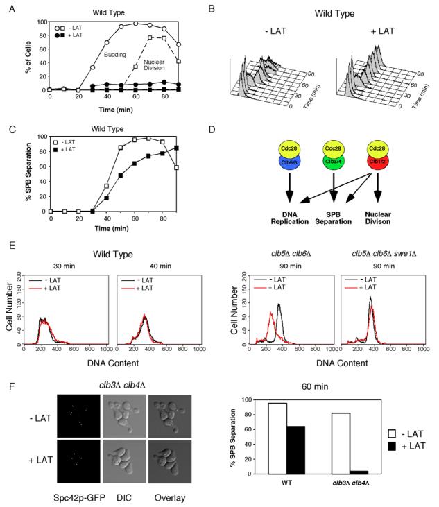
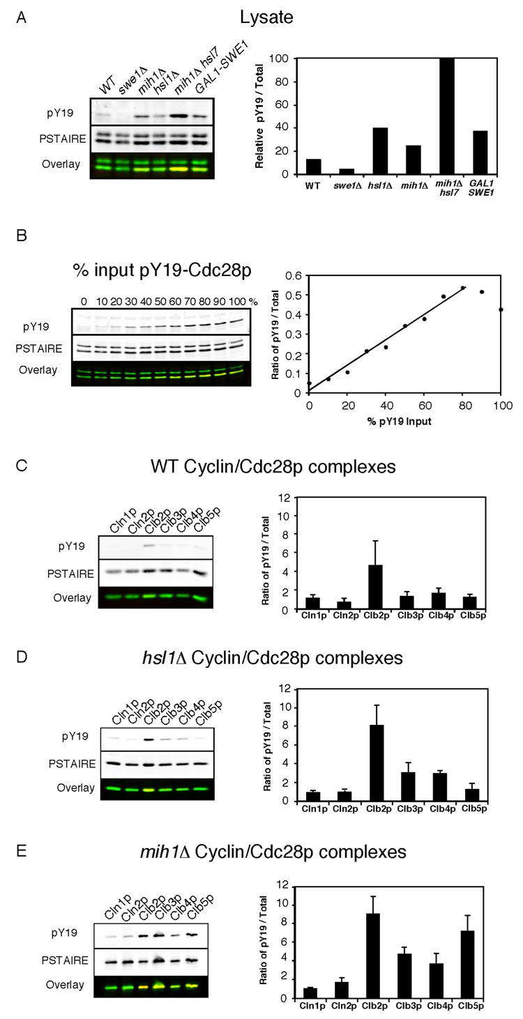
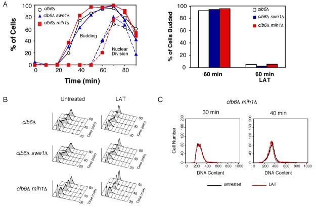
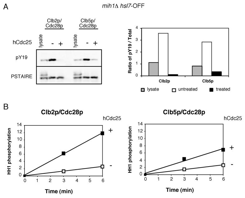
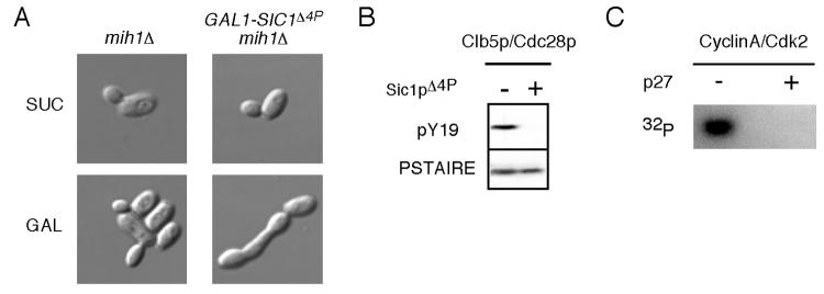
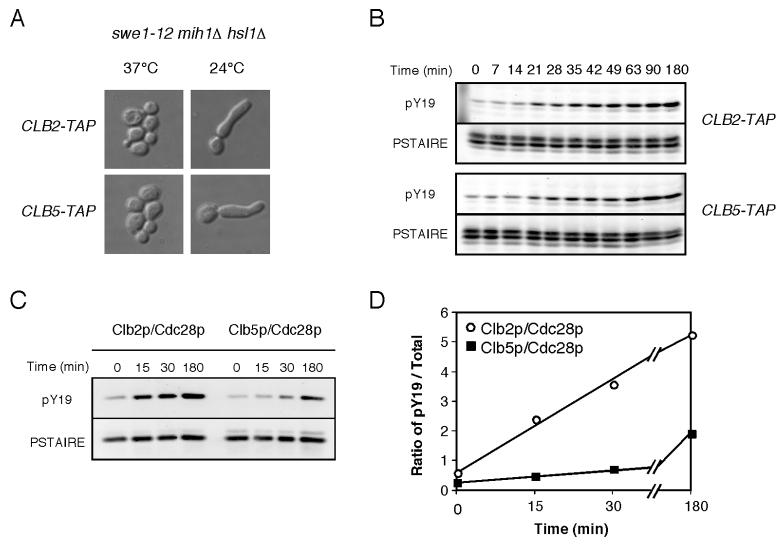
Results: We demonstrate that in Saccharomyces cerevisiae, several cyclin/CDK complexes are protected from inhibitory tyrosine phosphorylation, allowing Clb5,6p to promote DNA replication and Clb3,4p to promote spindle assembly, even under checkpoint-inducing conditions that block nuclear division. In vivo, S phase-promoting Clb5p/Cdc28p complexes were phosphorylated more slowly and dephosphorylated more effectively than were mitosis-promoting Clb2p/Cdc28p complexes. Moreover, we show that the CDK inhibitor (CKI) Sic1p protects bound Clb5p/Cdc28p complexes from tyrosine phosphorylation, allowing the accumulation of unphosphorylated complexes that are unleashed when Sic1p is degraded to promote S phase. The vertebrate CKI p27(Kip1) similarly protects Cyclin A/Cdk2 complexes from Wee1, suggesting that the antagonism between CKIs and Wee1 is evolutionarily conserved.

Conclusions: In yeast cells, the combination of CKI binding and preferential phosphorylation/dephosphorylation of different B cyclin/CDK complexes renders S phase progression immune from checkpoints acting via CDK tyrosine phosphorylation.


Figures

Figure 1
Figure 1. Effect of actin depolymerization on DNA replication, spindle assembly, and nuclear division
Cells were synchronized by pheromone arrest/release and part of the culture was treated with 150 μM LAT at 21 min after release from G1 arrest (just prior to budding). A) WT (DLY7974) cells were scored for budding (circles) and nuclear division (squares). The block of nuclear division was largely Swe1p dependent (Fig. S1A). B). LAT did not influence the timing of S phase, either in the presence or absence (Fig. S1B) of Swe1p as assessed by FACS analysis. C) Spindle formation was monitored by fluorescence microscopy to visualize the SPB marker Spc42-GFP. (n >200 cells). The small delay induced by LAT was largely Swe1p-dependent (Fig. S1C). D) Roles of different B-cyclin/Cdc28p complexes in the yeast cell cycle [18, 19, 21]. E) clb5Δ clb6Δ (DLY8300) and clb5Δ clb6Δ swe1Δ (DLY8963) cells were synchronized and DNA replication was monitored by flow cytometry (see Fig. S1D-E for synchrony data). Overlays of untreated (black) and LAT-treated (red) FACS profiles 90 min after release from G1 arrest. For comparison, WT FACS profiles during (30 min) and after (40 min) S phase are shown to illustrate lack of LAT effect in those cells (from data in B). F) WT (DLY7974) and clb3Δ clb4Δ (DLY8297) cells were synchronized and spindle formation was monitored as above (see Fig. S1G for synchrony data). Left, representative fluorescence and DIC images from clb3Δ clb4Δ cells. Right, >200 cells were scored for SPB separation at 60 min.
Figure 2
Figure 2. Selective phosphorylation of B cyclin/Cdc28p complexes
A) Y19 phosphorylation of the complete cellular pool of Cdc28p from WT (DLY7598), swe1Δ (DLY7712), hsl1Δ (DLY7944), mih1Δ (DLY7854), mih1Δ hsl7-OFF (DLY8197), and GAL1-SWE1 (DLY7946) strains. The first four strains were harvested during exponential growth, and the other strains were arrested by addition of dextrose (lane 5) or galactose (lane 6) as described in the Materials and Methods. The graph shows the ratio of phosphorylated (pY19)-to-total Cdc28p (PSTAIRE) for each strain, normalized to the level in mih1Δ hsl7-OFF arrested cells (this is the maximal experimentally obtained phosphorylation, but does not represent 100% phosphorylated Cdc28p because only B cyclin-bound Cdc28p is phosphorylated). Note that the anti-PSTAIRE antibody recognizes Pho85p (upper band) as well as Cdc28p (lower band) in whole cell lysates. B) Linearity of the Cdc28p phosphorylation measurement. Lysate from swe1Δ cells (DLY7712), lacking Cdc28p Y19 phosphorylation, was mixed with an increasing proportion of lysate from arrested mih1Δ hsl7-OFF cells (DLY8197), in which Cdc28p is maximally phosphorylated (the total amount of protein was equal in each lane). The ratio of pY19-to-total Cdc28p signal was plotted against the percentage of input lysate from the mih1Δ hsl7-OFF strain. C-E) The indicated cyclin/Cdc28p complexes were isolated from the following strains harvested during exponential growth: C) WT (DLY7646, DLY7647, DLY7598, DLY7648, DLY7599, DLY7662); D) hsl1Δ (DLY7735, DLY7718, DLY7873 DLY7770, DLY7749); E) mih1Δ (DLY7857, DLY7717, DLY7854, DLY7858, DLY7860, DLY7786). The ratio of pY19-to-total Cdc28p was quantitated for both the purified complexes and the complete cellular pool of Cdc28p in lysates for each experiment. To account for gel-to-gel variability, the results from individual experiments were normalized by dividing the ratio for each purified complex by the average ratio obtained from lysates used in that experiment. The average normalized ratios for each cyclin/Cdc28p complex are displayed in arbitrary units as mean +/− SEM (n = 3). (pY19, red; PSTAIRE, green)
Figure 3
Figure 3. LAT does not affect DNA replication even in the absence of Mih1p
Cells were synchronized as in Fig. 1. A) Budding and nuclear division in clb6Δ (DLY8245 ○), clb6Δ swe1Δ (DLY8261, ), and clb6Δ mih1Δ (DLY8441, ) cells were monitored by fluorescence microscopy (n >200). B) DNA replication was monitored by flow cytometry. C) FACS profiles for untreated (black) and LAT treated (red) clb6Δ mih1Δ cells 30 min and 40 min after release from G1 arrest.
Figure 4
Figure 4. Tyrosine phosphorylation inhibits both Clb5p- and Clb2p-associated kinase activity
A) mih1Δ GAL1-HSL7 cells (DLY8197 and DLY8199) were grown in dextrose-containing medium for 22 h to deplete Hsl7p (mih1Δ hsl7-OFF). Clb2p/Cdc28p and Clb5p/Cdc28p complexes were isolated, treated with or without recombinant hCdc25, and the ratio of pY19-to-total Cdc28p was assessed. B) Phosphorylated (mock treated, □) and dephosphorylated (hCDC25 treated, ) Cdc28p complexes were assayed for their ability to phosphorylate histone H1 in vitro. The amount of 32P incorporation was normalized to the amount of Cdc28p present in the reaction and is plotted in arbitrary units (mean of two replicates).
Figure 5
Figure 5. CKIs protect bound CDK from tyrosine phosphorylation
A) Representative DIC images of mih1Δ (DLY8700) and GAL1-SIC1Δ4p mih1Δ (DLY8701) cells grown in YEPS (SUC) and induced to express Sic1p by addition of galactose for 5 h (GAL). B) Cells from the same strains were grown in YEPS and arrested in G1 with pheromone. Sic1pΔ4p expression was induced with galactose 30 min prior to release from the G1 arrest and maintained following release into YEPG. Cdc28p Y19 phosphorylation was assessed for Clb5p/Cdc28p complexes isolated from cells collected 75 min after release when the majority of the cells had budded. C) Purified Cyclin A/Cdk2 was incubated with GST-Wee1 and γ-32P-ATP in the presence or absence of p27Kip1 as described in the Materials and Methods. Phosphorylated Cdk2 was visualized using a phosphoimager.
Figure 6
Figure 6. Swe1p phosphorylates Clb2p/Cdc28p more rapidly than Clb5p/Cdc28p
A) swe1-12 hsl1Δ mih1Δ cells containing Clb2p-TAP (DLY8726) or Clb5p-TAP (DLY8727) were grown at 37°C and shifted to 24°C for 5 h and processed for DIC imaging. B) Cells from those strains were grown in 0.5 M HU at 37°C for 5 h and shifted to 24°C in the continued presence of HU (>90% large-budded arrest). Cdc28p phosphorylation was assessed by Western blot analysis of TCA-precipitated total cellular protein. C) Clb2p and Clb5p complexes were isolated from cells in B at the indicated times after shift to 24°C, and Cdc28p Y19 phosphorylation was assessed. D) The ratio of pY19-to-total Cdc28p was determined for the purified Cdc28p complexes shown in C.

References

    1. Morgan DO. Principles of CDK regulation. Nature. 1995;374:131–134. - PubMed
    1. Humphrey T. DNA damage and cell cycle control in Schizosaccharomyces pombe. Mutat. Res. 2000;451:211–226. - PubMed
    1. Donzelli M, Draetta GF. Regulating mammalian checkpoints through Cdc25 inactivation. EMBO Rep. 2003;4:671–677. - PMC - PubMed
    1. Lew DJ. The morphogenesis checkpoint: how yeast cells watch their figures. Curr. Opin. Cell Biol. 2003;15:648–653. - PubMed
    1. Falck J, Mailand N, Syljuasen RG, Bartek J, Lukas J. The ATM-Chk2-Cdc25A checkpoint pathway guards against radioresistant DNA synthesis. Nature. 2001;410:842–847. - PubMed

Publication types

MeSH terms