

Regulation of ligands for the activating receptor NKG2D
- PMID: 17614877
- PMCID: PMC2265965
- DOI: 10.1111/j.1365-2567.2007.02652.x
Regulation of ligands for the activating receptor NKG2D
Abstract
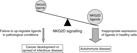
The outcome of an encounter between a cytotoxic cell and a potential target cell depends on the balance of signals from inhibitory and activating receptors. Natural Killer group 2D (NKG2D) has recently emerged as a major activating receptor on T lymphocytes and natural killer cells. In both humans and mice, multiple different genes encode ligands for NKG2D, and these ligands are non-classical major histocompatibility complex class I molecules. The NKG2D-ligand interaction triggers an activating signal in the cell expressing NKG2D and this promotes cytotoxic lysis of the cell expressing the ligand. Most normal tissues do not express ligands for NKG2D, but ligand expression has been documented in tumour and virus-infected cells, leading to lysis of these cells. Tight regulation of ligand expression is important. If there is inappropriate expression in normal tissues, this will favour autoimmune processes, whilst failure to up-regulate the ligands in pathological conditions would favour cancer development or dissemination of intracellular infection.
Figures


Similar articles
-
NKG2D receptor-mediated NK cell function is regulated by inhibitory Ly49 receptors.Blood. 2005 Jan 1;105(1):233-40. doi: 10.1182/blood-2004-03-1075. Epub 2004 Aug 24. Blood. 2005. PMID: 15328154 Free PMC article.
-
Receptors and lytic mediators regulating anti-tumor activity by the leukemic killer T cell line TALL-104.J Leukoc Biol. 2005 Aug;78(2):359-71. doi: 10.1189/jlb.0604360. Epub 2005 Jun 3. J Leukoc Biol. 2005. PMID: 15937142
-
NKG2D triggering hampers DNAM-1-mediated signaling in human NK cells.Front Immunol. 2025 May 12;16:1575059. doi: 10.3389/fimmu.2025.1575059. eCollection 2025. Front Immunol. 2025. PMID: 40421025 Free PMC article.
-
The NKG2D receptor: sensing stressed cells.Trends Mol Med. 2008 Apr;14(4):179-89. doi: 10.1016/j.molmed.2008.02.004. Epub 2008 Mar 18. Trends Mol Med. 2008. PMID: 18353724 Review.
-
NKG2D ligands: key targets of the immune response.Trends Immunol. 2008 Aug;29(8):397-403. doi: 10.1016/j.it.2008.04.007. Epub 2008 Jul 3. Trends Immunol. 2008. PMID: 18602338 Review.
Cited by
-
Soluble ligands for the NKG2D receptor are released during endometriosis and correlate with disease severity.PLoS One. 2015 Mar 16;10(3):e0119961. doi: 10.1371/journal.pone.0119961. eCollection 2015. PLoS One. 2015. PMID: 25775242 Free PMC article.
-
Which NK cell populations mark the high burden of CMV present in all HIV patients beginning ART in Indonesia?AIDS Res Ther. 2022 Mar 15;19(1):16. doi: 10.1186/s12981-022-00439-2. AIDS Res Ther. 2022. PMID: 35292053 Free PMC article.
-
Natural killer cell receptors and their ligands in liver diseases.Med Mol Morphol. 2009 Mar;42(1):1-8. doi: 10.1007/s00795-008-0434-7. Epub 2009 Mar 18. Med Mol Morphol. 2009. PMID: 19294486 Review.
-
Design and Implementation of NK Cell-Based Immunotherapy to Overcome the Solid Tumor Microenvironment.Cancers (Basel). 2020 Dec 21;12(12):3871. doi: 10.3390/cancers12123871. Cancers (Basel). 2020. PMID: 33371456 Free PMC article. Review.
-
Chemotherapy-resistant osteosarcoma is highly susceptible to IL-15-activated allogeneic and autologous NK cells.Cancer Immunol Immunother. 2011 Apr;60(4):575-86. doi: 10.1007/s00262-010-0965-3. Epub 2011 Jan 15. Cancer Immunol Immunother. 2011. PMID: 21240486 Free PMC article.
References
-
- Moretta A, Bottino C, Vitale M, Pende D, Cantoni C, Mingari MC, Biassoni R, Moretta L. Activating receptors and coreceptors involved in human natural killer cell-mediated cytolysis. Annu Rev Immunol. 2001;19:197–223. - PubMed
-
- Braud VM, Allan DS, O'Callaghan CA, et al. HLA-E binds to natural killer cell receptors CD94/NKG2A, B and C. Nature. 1998;391:795–9. - PubMed
-
- O'Callaghan CA. Molecular basis of human natural killer cell recognition of HLA-E (human leucocyte antigen-E) and its relevance to clearance of pathogen-infected and tumour cells. Clin Sci (Lond) 2000;99:9–17. - PubMed
-
- O'Callaghan CA. Natural killer cell surveillance of intracellular antigen processing pathways mediated by recognition of HLA-E and Qa-1b by CD94/NKG2 receptors. Microbes Infect. 2000;2:371–80. - PubMed
-
- Karre K, Ljunggren HG, Piontek G, Kiessling R. Selective rejection of H-2-deficient lymphoma variants suggests alternative immune defence strategy. Nature. 1986;319:675–8. - PubMed
Publication types
MeSH terms
Substances
Grants and funding
LinkOut - more resources
Full Text Sources
Other Literature Sources
