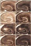
Acute and chronic changes in glycogen phosphorylase in hippocampus and entorhinal cortex after status epilepticus in the adult male rat
- PMID: 17614948
- PMCID: PMC2504499
- DOI: 10.1111/j.1460-9568.2007.05657.x
Acute and chronic changes in glycogen phosphorylase in hippocampus and entorhinal cortex after status epilepticus in the adult male rat
Abstract
Glial cells provide energy substrates to neurons, in part from glycogen metabolism, which is influenced by glycogen phosphorylase (GP). To gain insight into the potential subfield and laminar-specific expression of GP, histochemistry can be used to evaluate active GP (GPa) or totalGP (GPa + GPb). Using this approach, we tested the hypothesis that changes in GP would occur under pathological conditions that are associated with increased energy demand, i.e. severe seizures (status epilepticus or 'status'). We also hypothesized that GP histochemistry would provide insight into changes in the days and weeks after status, particularly in the hippocampus and entorhinal cortex, where there are robust changes in structure and function. One hour after the onset of pilocarpine-induced status, GPa staining was reduced in most regions of the hippocampus and entorhinal cortex relative to saline-injected controls. One week after status, there was increased GPa and totalGP, especially in the inner molecular layer, where synaptic reorganization of granule cell mossy fibre axons occurs (mossy fibre sprouting). In addition, patches of dense GP reactivity were evident in many areas. One month after status, levels of GPa and totalGP remained elevated in some areas, suggesting an ongoing role of GP or other aspects of glycogen metabolism, possibly due to the evolution of intermittent, recurrent seizures at approximately 3-4 weeks after status. Taken together, the results suggest that GP is dynamically regulated during and after status in the adult rat, and may have an important role in the pilocarpine model of epilepsy.
Figures


Similar articles
-
Neuronal degeneration is observed in multiple regions outside the hippocampus after lithium pilocarpine-induced status epilepticus in the immature rat.Neuroscience. 2013 Nov 12;252:45-59. doi: 10.1016/j.neuroscience.2013.07.045. Epub 2013 Jul 27. Neuroscience. 2013. PMID: 23896573 Free PMC article.
-
Glycogen phosphorylase reactivity in the entorhinal complex in familiar and novel environments: evidence for labile glycogenolytic modules in the rat.J Chem Neuroanat. 2006 Feb;31(2):108-13. doi: 10.1016/j.jchemneu.2005.09.004. Epub 2005 Oct 17. J Chem Neuroanat. 2006. PMID: 16229987
-
Long-term pregabalin treatment protects basal cortices and delays the occurrence of spontaneous seizures in the lithium-pilocarpine model in the rat.Epilepsia. 2003 Jul;44(7):893-903. doi: 10.1046/j.1528-1157.2003.61802.x. Epilepsia. 2003. PMID: 12823571
-
Recurrent mossy fiber pathway in rat dentate gyrus: synaptic currents evoked in presence and absence of seizure-induced growth.J Neurophysiol. 1999 Apr;81(4):1645-60. doi: 10.1152/jn.1999.81.4.1645. J Neurophysiol. 1999. PMID: 10200201
-
Early Aberrant Growth of Mossy Fibers after Status Epilepticus in the Immature Rat Brain.Mol Neurobiol. 2019 Jul;56(7):5025-5031. doi: 10.1007/s12035-018-1432-y. Epub 2018 Nov 17. Mol Neurobiol. 2019. PMID: 30448889
Cited by
-
Modulation of the perforant path-evoked potential in dentate gyrus as a function of intrahippocampal β-adrenoceptor agonist concentration in urethane-anesthetized rat.Brain Behav. 2014 Jan;4(1):95-103. doi: 10.1002/brb3.199. Epub 2013 Dec 20. Brain Behav. 2014. PMID: 24653959 Free PMC article.
-
Effect of eugenol on lithium-pilocarpine model of epilepsy: behavioral, histological, and molecular changes.Iran J Basic Med Sci. 2017 Jul;20(7):745-752. doi: 10.22038/IJBMS.2017.9004. Iran J Basic Med Sci. 2017. PMID: 28852438 Free PMC article.
-
Glycogen serves as an energy source that maintains astrocyte cell proliferation in the neonatal telencephalon.J Cereb Blood Flow Metab. 2017 Jun;37(6):2294-2307. doi: 10.1177/0271678X16665380. Epub 2016 Jan 1. J Cereb Blood Flow Metab. 2017. PMID: 27601444 Free PMC article.
-
Brain glycogen content is increased in the acute and interictal chronic stages of the mouse pilocarpine model of epilepsy.Epilepsia Open. 2022 Jun;7(2):361-367. doi: 10.1002/epi4.12599. Epub 2022 Apr 22. Epilepsia Open. 2022. PMID: 35377551 Free PMC article.
-
Does abnormal glycogen structure contribute to increased susceptibility to seizures in epilepsy?Metab Brain Dis. 2015 Feb;30(1):307-16. doi: 10.1007/s11011-014-9524-5. Epub 2014 Mar 19. Metab Brain Dis. 2015. PMID: 24643875 Free PMC article. Review.
References
-
- Bernard-Helary K, Lapouble E, Ardourel M, Hevor T, Cloix JF. Correlation between brain glycogen and convulsive state in mice submitted to methionine sulfoximine. Life Sci. 2000;67:1773–1781. - PubMed
-
- Bolwig TG, Rafaelsen OJ. Brain glycogen and serum glucose after convulsions induced by electroshock and pentamenthylenetetrazole in rats. Acta Psychiatr Scand. 1972;48:377–385. - PubMed
-
- Borges K, Gearing M, McDermott DL, Smith AB, Almonte AG, Wainer BH, Dingledine R. Neuronal and glial pathological changes during epileptogenesis in the mouse pilocarpine model. Exp Neurol. 2003;182:21–34. - PubMed
-
- Brown AM, Sickmann HM, Fosgerau K, Lund TM, Schousboe A. Astrocyte glycogen metabolism is required for neural activity during aglycemia or intense stimulation in mouse white matter. J Neurosci Res. 2005;79:74–80. - PubMed
-
- Brushia RJ, Walsh DA. Phosphorylase kinase: the complexity of its regulation is reflected in the complexity of its structure. Front Biosci. 1999;4:D618–D641. - PubMed
Publication types
MeSH terms
Substances
Grants and funding
LinkOut - more resources
Full Text Sources

