. 2007 Sep 1;583(Pt 2):593-609.
doi: 10.1113/jphysiol.2007.135947. Epub 2007 Jul 5.

Phenotypic changes of morphologically identified guinea-pig myenteric neurons following intestinal inflammation

Affiliations

Phenotypic changes of morphologically identified guinea-pig myenteric neurons following intestinal inflammation

Kulmira Nurgali et al. J Physiol. .

Abstract

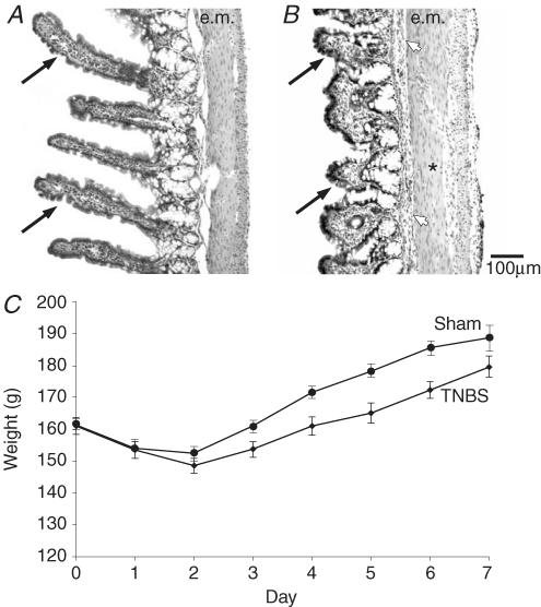
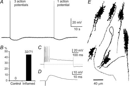
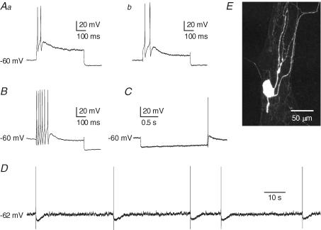
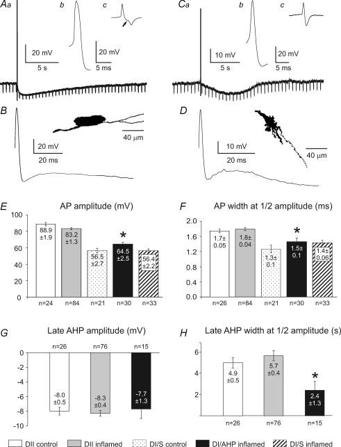
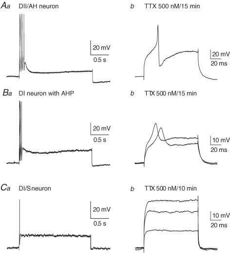
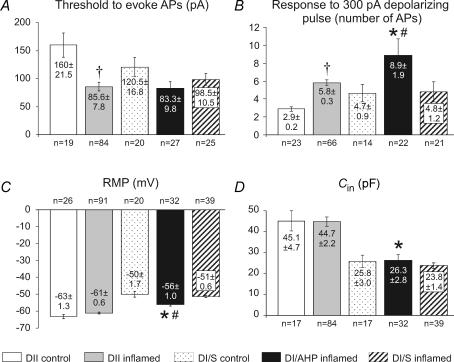
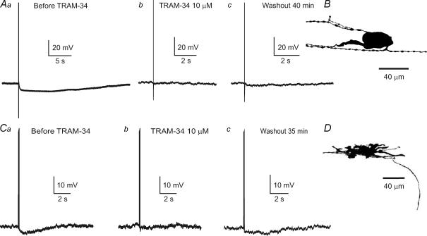
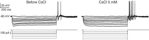
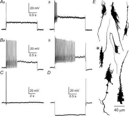
We investigated the responses of morphologically identified myenteric neurons of the guinea-pig ileum to inflammation that was induced by the intraluminal injection of trinitrobenzene sulphonate, 6 or 7 days previously. Electrophysiological properties were examined with intracellular microelectrodes using in vitro preparations from the inflamed or control ileum. The neurons were injected with marker dyes during recording and later they were recovered for morphological examination. A proportion of neurons with Dogiel type I morphology, 45% (32/71), from the inflamed ileum had a changed phenotype. These neurons exhibited an action potential with a tetrodotoxin-resistant component, and a prolonged after-hyperpolarizing potential followed the action potential. Of the other 39 Dogiel type I neurons, no changes were observed in 36 and 3 had increased excitability. The afterhyperpolarizing potential (AHP) in Dogiel type I neurons was blocked by the intermediate conductance, Ca(2+)-dependent K(+) channel blocker TRAM-34. Neurons which showed these phenotypic changes had anally directed axonal projections. Neither a tetrodotoxin-resistant action potential nor an AHP was seen in Dogiel type I neurons from control preparations. Dogiel type II neurons retained their distinguishing AH phenotype, including an inflection on the falling phase of the action potential, an AHP and, in over 90% of neurons, an absence of fast excitatory transmission. However, they became hyperexcitable and exhibited anodal break action potentials, which, unlike control Dogiel type II neurons, were not all blocked by the h current (I(h)) antagonist Cs(+). It is concluded that inflammation selectively affects different classes of myenteric neurons and causes specific changes in their electrophysiological properties.


Figures

Figure 1
Figure 1. Changes indicative of inflammation: reduced villus height and body weight loss
A and B, haematoxylin and eosin staining of the ileum from control (A), and from a guinea-pig 6 days after the intraluminal injection of 2,4,6-trinitrobenzenesulphonic acid (TNBS, B). Comparison of the images shows the substantial decrease in the heights of villi after TNBS (arrows). There is also a slight thickening of the external muscle (e.m.) and enlarged lymphatic vessels (*). The submucosal layer is more prominent (arrowhead). C, time course of body weight loss and recovery after injection of TNBS into the guinea-pig ileum (♦, mean ±s.e.m., n = 155). Changes in body weight in sham controls reveal a smaller weight loss and quicker post-operative weight gain (•, n = 9).
Figure 2
Figure 2. The occurrence of afterhyperpolarizing potentials (AHPs) in myenteric neurons with Dogiel type I (DI) morphology from the inflamed ileum
A, examples of late AHPs in a DI neuron. A train of three action potentials as well as a single action potential, elicited by brief intracellular current pulses, are followed by late AHPs lasting about 8 s. B, late AHPs did not occur in these neurons from control ileum (0/22), whereas about 45% (32/71) of DI neurons from the inflamed ileum exhibited late AHPs. C, a depolarizing current pulse of 100 pA and 500 ms induced 7 action potentials. Early AHPs, lasting less than 10 ms, are seen after each action potential. Following the action potential burst, the development of the late AHP is apparent as a sag in the depolarization and a hyperpolarization of about 15 mV after the end of current injection. D, a fast excitatory postsynaptic potential elicited by stimulation of presynaptic inputs to a DI neuron with a late AHP. Fast EPSPs were prominent in all DI neurons with late AHPs. E, examples of the shapes of DI neurons that exhibited late AHPs. Each of these neurons is from the inflamed intestine and was filled with dye during recording. The typical short lamellar dendrites and single axon, sometimes with axonal spines, of DI neurons are seen.
Figure 7
Figure 7. Action potential generation in Dogiel type II (DII) neurons from the inflamed ileum
Aa and b, action potential firing in response to depolarizing current pulse (200 pA, 500 ms) of DII neurons from control (Aa) and sham-operated (Ab) ileum. B, action potentials in response to intracellular depolarizing current (200 pA, 500 ms) in a DII neuron from the inflamed ileum. C, anodal break action potential that was observed in a DII neuron from the inflamed intestine. Anodal break action potentials were much more common after inflammation. D, spontaneous action potentials arising at a resting membrane potential of −62 mV in a DII neuron from the inflamed intestine. Spontaneous action potentials were never seen in Dogiel type II neurons from control intestine, under the conditions used in this study, even when the resting membrane potential was more depolarized than −62 mV. E, fluorescence image of the neuron from which the record in B was taken. This is a typical DII neuron with a smooth-surfaced oval cell body and several long, axon-like processes.
Figure 3
Figure 3. Properties of afterhyperpolarizing potentials in myenteric Dogiel type II (DII) and Dogiel type I (DI) neurons from the inflamed ileum
Aa, b and c, late AHP and action potential in a DII neuron. The AHP following a single action potential was associated with significant reduction of the input resistance, as indicated by the decrease in the voltage changes caused by hyperpolarizing current injection (Aa). The action potential had a hump on the falling phase (Ab), demonstrated in the first time derivative (dV/dt) of the action potential (Ac), which clearly shows the biphasic falling phase (arrow). B, the action potential and after-potentials of the neuron in A at greater sweep speed. It can be seen that the membrane potential depolarizes after the early AHP, before the beginning of the late AHP is apparent. The inset shows the shape of the neuron from which the records in A were taken. This is a typical DII neuron, with an oval cell body and multiple long processes (camera lucida drawing after dye filling). Ca, b and c, the action potential and afterhyperpolarization in a DI neuron. The late AHP following a single action potential was also associated with a reduction in input resistance (Ca). In both cells the action potential was recorded with the membrane potential held at −60 mV. The action potential did not have a hump on its falling phase (Cb), which is emphasized by the lack of inflection in the first derivative of the trace (Cc). D, the action potential and after potentials, showing the delay before the onset of the late AHP. The inset shows the neuron from which the records in C and D were taken. This is a typical DI neuron, with lamellar dendrites and a single axon. E and F, action potential (AP) amplitudes and widths at half-amplitude of the APs of DII neurons were not significantly affected by inflammation. G, amplitudes of late AHPs of DII neurons from control and inflamed ileum and DI neurons from the inflamed ileum. There are no significant differences. H, widths at half-amplitude of late AHPs of DII neurons from control and inflamed ileum and DI neurons from the inflamed ileum. The late AHP of the DI neurons was briefer than that of DII neurons (P < 0.05). Means ± s.e.m. and numbers of neurons analysed for each parameter are given in the histograms. *Significantly different from Dogiel type II neurons from the inflamed ileum (P < 0.05).
Figure 4
Figure 4. Sensitivities to tetrodotoxin (TTX) of action potentials of Dogiel type II (DII) and Dogiel type I (DI) neurons from the inflamed ileum
Aa and b, action potentials in response to intracellular depolarizing current (200 pA, 2 s) in a DII neuron from the inflamed ileum (A) and in the same neuron 15 min after exposure to TTX (500 nm). Depolarization (300 pA, 100 ms) still elicited an active event, although its amplitude was reduced (Ab). Ba and b, for a DI neuron from the inflamed ileum, in which late AHPs were observed, the action potential induced by a 200 pA, 2 s depolarizing pulse in control solution (Ba) was reduced but not blocked by TTX (Bb, 200 and 250 pA, 100 ms pulses). Ca and b, the action potential in DI neurons without late AHPs from the inflamed ileum in response to intracellular depolarizing current (200 pA, 2 s, Ca) was completely blocked by TTX (Cb, 150, 250 and 300 pA, 100 ms pulses). In all cases the membrane potential prior to depolarization was held at −60 mV.
Figure 5
Figure 5. Comparisons of electrophysiological properties of Dogiel type II (DII) and Dogiel type I (DI) neurons from the control and inflamed ileum
Five groups are compared: DII neurons from control and inflamed ileum, DI neurons from control, DI neurons that exhibited late AHPs from the inflamed ileum and DI neurons that did not exhibit late AHPs from the inflamed ileum. A, thresholds for action potential generation in DII neurons from the inflamed ileum were significantly lower than from control (P < 0.001). Differences between DI neurons were not significant. B, excitability, measured as the numbers of action potentials elicited by 500 ms depolarizing pulses, was significantly greater for DI/AHP neurons from the inflamed intestine compared with DI neurons from control or DI/S neurons from the inflamed ileum (P < 0.05). Excitability was also increased for DII neurons from the inflamed ileum compared with DII neurons from the control ileum (P < 0.05). C, the resting membrane potential (RMP) was not changed in DII neurons, although DI/AHP neurons from the inflamed ileum were more hyperpolarized than DI/S inflamed or control DI neurons (P < 0.05), but less hyperpolarized than DII neurons from the inflamed ileum (P < 0.001). D, there were no significant differences in neuron capacitance. DI/AHP neurons from the inflamed intestine were significantly smaller than DII neurons. Values of mean ± s.e.m. and numbers of neurons analysed for each parameter are incorporated into the histograms. *Significantly different from Dogiel type II neurons from the inflamed ileum (P < 0.05). #Significantly different from Dogiel type I/S neurons from the inflamed ileum (P < 0.05). †Significantly different from Dogiel type II neurons from the control ileum (P < 0.05).
Figure 6
Figure 6. Inhibition of late AHPs by a blocker of intermediate conductance Ca2+-activated potassium (IKCa) channels, TRAM-34
Aa, b and c, late AHP in a Dogiel type II neuron from the inflamed intestine before the (Aa) and after application of TRAM-34 (10 μm, Ab). The amplitude of late AHP did not recover even after 40 min of washout with Krebs solution (Ac). B, the shape of the neuron from which the records in A were taken. Ca, b and c, late AHP in a Dogiel type I neuron was also blocked by TRAM-34 (10 μm). D, the shape of the Dogiel type I neuron with a late AHP from which the records in C were taken. The membrane potential in both neurons was held at −60 mV.
Figure 8
Figure 8. Persistence of anodal break action potentials in DII neurons from the inflamed ileum after additin of Cs+
In control solution there was an obvious sag in the voltage response to imposed hyperpolarization, which is due to the activation of a hyperpolarization activated cation current (Ih). Ih persists after the end of hyperpolarizing steps, and this tail current has been implicated as a cause of the anodal break. However, when Ih was blocked by Cs+ (5 mm), anodal break action potentials were still observed in some DII neurons from the inflamed intestine.
Figure 9
Figure 9. Excitability and morphology of Dogiel type I neurons without AHPs from the inflamed ileum
Aa and b, excitability of a DI neuron with typical S electrophysiological properties (no AHP) from the inflamed ileum. Such neurons were 25/28 DI/S neurons that were tested. The threshold for depolarization to evoke an action potential was about 100 pA (Aa). The number of action potentials in response to a 300 pA depolarizing pulse was up to six (Ab). Ba and b, records from a hyperexcitable DI/S neuron from the inflamed ileum. These were 3/28 of the DI/S neurons. B, the hyperexcitable neurons responded with multiple action potentials to a 100 pA depolarizing current pulse. Bb, response to 300 pA depolarizing current pulses revealed their hyperexcitability. C, the three hyperexcitable neurons did not have a late AHP, but had prominent anodal break action potentials (D). Shapes of DI/S neurons without AHPs from the inflamed intestine are shown at the right (E). The records in Ba and b were from the neuron marked with an asterisk. Holding potential was −50 mV in A, Aa, B, Ba and D.

Similar articles

Cited by

References

    1. Beyak MJ, Ramji N, Krol KM, Kawaja MD, Vanner SJ. Two TTX-resistant Na+ currents in mouse colonic dorsal root ganglia neurons and their role in colitis-induced hyperexcitability. Am J Physiol Gastrointest Liver Physiol. 2004;287:G845–G855. - PubMed
    1. Bielefeldt K, Ozaki N, Gebhart GF. Mild gastritis alters voltage-sensitive sodium currents in gastric sensory neurons in rats. Gastroenterology. 2002;122:752–761. - PubMed
    1. Black JA, Liu S, Tanaka M, Cummins TR, Waxman SG. Changes in the expression of tetrodotoxin-sensitive sodium channels within dorsal root ganglia neurons in inflammatory pain. Pain. 2004;108:237–247. - PubMed
    1. Brehmer A, Schrodl F, Neuhuber W. Morphological classifications of enteric neurons – 100 years after Dogiel. Anat Embryol. 1999;200:125–135. - PubMed
    1. Brookes SJH. Classes of enteric nerve cells in the guinea-pig small intestine. Anat Rec. 2001;262:58–70. - PubMed

Publication types

MeSH terms

LinkOut - more resources