. 2007 Sep;18(9):3512-22.
doi: 10.1091/mbc.e07-04-0306. Epub 2007 Jul 5.

Serine phosphorylation of the integrin beta4 subunit is necessary for epidermal growth factor receptor induced hemidesmosome disruption

Affiliations

Serine phosphorylation of the integrin beta4 subunit is necessary for epidermal growth factor receptor induced hemidesmosome disruption

Kevin Wilhelmsen et al. Mol Biol Cell. 2007 Sep.

Abstract

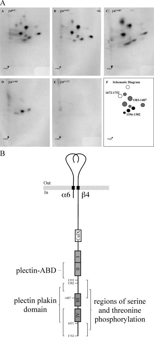
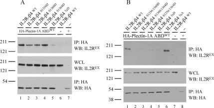
Hemidesmosomes (HDs) are multiprotein adhesion complexes that promote attachment of epithelial cells to the basement membrane. The binding of alpha6beta4 to plectin plays a central role in their assembly. We have defined three regions on beta4 that together harbor all the serine and threonine phosphorylation sites and show that three serines (S1356, S1360, and S1364), previously implicated in HD regulation, prevent the interaction of beta4 with the plectin actin-binding domain when phosphorylated. We have also established that epidermal growth factor receptor activation, which is known to function upstream of HD disassembly, results in the phosphorylation of only one or more of these three residues and the partial disassembly of HDs in keratinocytes. Additionally, we show that S1360 and S1364 of beta4 are the only residues phosphorylated by PKC and PKA in cells, respectively. Taken together, our studies indicate that multiple kinases act in concert to breakdown the structural integrity of HDs in keratinocytes, which is primarily achieved through the phosphorylation of S1356, S1360, and S1364 on the beta4 subunit.


Figures

Figure 1.
Figure 1.
Serine/threonine phosphorylation of β4 occurs predominantly in the CS and the C-terminal tail. (A) In vivo phosphopeptide maps of wild type (A) and four sequentially truncated (B–E) β4 subunits derived from COS-7 cells that were treated with calyculin A in the presence of [32P]orthophosphate. (F) A schematic diagram of the results shown in A–E. (B) Schematic drawings of the integrin α6β4 depicting the regions serine and threonine phosphorylated on the β4 subunit after calyculin A treatment and the interaction sites with plectin reported in the literature. Residue numbers indicate where the β4 deletions were made. Roman numerals I–IV show the positions of the four FnIII domains. The CS is between FnIII II and III, whereas the C-terminal tail is after FnIII IV. CalX, Ca-Na exchanger motif. β4 numbering is based on the human β4A sequence.
Figure 2.
Figure 2.
Phosphomimick aspartic acid substitutions of S1356, S1360, and S1364 in the β4 CS prevents the association with the Plectin-1A ABD. (A) COS-7 cells were transiently transfected with either IL2R-β4WT (lanes 1 and 6), IL2R-β4S1356D (lane 2), IL2R-β4S1360D (lane 3), IL2R-β4S1364D (lane 4), or IL2R-β43xD (lane 5) cDNA constructs or a control plasmid (lane 7) and an expression construct for the HA-plectin-1A ABDWT (lanes 1–5 and 7) or a control plasmid (lane 6). The cells were lysed in M-PER buffer and HA-IPs were probed for the presence of IL2R-β4 (top) and the HA-tagged ABDs (bottom). Whole cell lysates (WCLs) were probed for the expression level of IL2R-β4 proteins (middle). (B) The experiment was performed as in A, except that either IL2R-β4WT (lanes 1 and 7), IL2R-β4S1356/1360D (lane 2), IL2R-β4S1360/1364D (lane3), IL2R-β4S1356/1364D (lane 4), IL2R-β43xD (lane 5), or IL2R-β43xA (lane 6) cDNA constructs, or a control plasmid (lane 8), were instead cotransfected with the HA-tagged plectin-1A ABD cDNA construct (lanes 1–6 and 8) or a control plasmid (lane 7).
Figure 3.
Figure 3.
S1360 and S1364 are in vivo PKC and PKA phosphorylation sites on β4, respectively, that cooperate to reduce the association with the plectin-1A ABD. (A) In vivo phosphopeptide maps of β4 isolated from either PA-JEB/β4WT cells that were serum-starved (A), treated with PMA (B) or calyculin A (D), PA-JEB/β4S1360A cells that were treated with PMA (C) or PA-JEB/β43xA cells that were treated with calyculin A (E) in the presence of [32P]orthophosphate. (F) A schematic diagram of the results shown in A–E. Gray ovals, peptides containing β4 specific phosphorylation sites. (B) PA-JEB/β4WT cells were either left serum-starved (lane 1), treated with PMA for 30 min (lane 2), or treated with PMA for 30 min in the presence of the compound Gö6983 (lane 3) before lysis in mPER buffer. Similarly, PA-JEB/β43xA (lanes 4 and 5) and PA-JEB/β43xD (lanes 6 and 7) cells were either left serum-starved (lanes 4 and 6) or treated with PMA for 30 min (lanes 5 and 7) before lysis. WCLs were probed for the presence of β4 phosphorylated on residues S1356, S1360, and/or S1364 using our rabbit polyclonal phospho-specific antibody β4 pS-CS (top). The expression level of the β4 proteins was detected using a rabbit polyclonal antibody raised against the first pair of FNIII repeats in β4 (middle). To verify the activation of PKC, WCLs were also probed for the presence of phospho-PKC using pan antibodies raised against phosphorylated Thr 660 of PKCβ II (bottom). (C) Alignment of the human β4A sequence from residue 1352-1368 with the same region in cow, dog, mouse, rat, chicken, and zebrafish. The kinases identified from the phospho-phosphorylation site prediction programs phospho-ELM and GPS are shown below their corresponding residue. The human β4 sequence was used as the input. (D) In vivo phosphopeptide maps of β4 isolated from either serum-starved PA-JEB/β4WT cells (A) or PA-JEB/β4WT (B), PA-JEB/β4S1360/1364A (C) or PA-JEB (an equivalent area of gel were β4 would run was excised; D) cells treated for 30 min with FSK/IBMX in the presence of [32P]orthophosphate. (E) A schematic diagram of the results shown in A–D. Note that the peptides in these maps migrate to a lower part on the thin-layer chromatography plates because the chromatography buffer used was not optimal. Gray ovals, peptides containing β4 specific phosphorylation sites. (E) PA-JEB/β4WT cells were either serum-starved (lane 1) or treated with Forskolin/IBMX for the indicated times (in minutes; lanes 2–7) before lysis in mPER buffer. WCLs were probed for the presence of β4 phosphorylated on residues S1356, S1360, and/or S1364 using our rabbit polyclonal phospho-specific antibody β4 pS-CS (top) and the expression level of the β4 proteins with our rabbit polyclonal antibody raised against the first pair of FNIII repeats in β4 (bottom). (F) COS-7 cells were transiently transfected with either IL2R-β4WT (lanes 1–4 and 7) or IL2R-β4S1360/1364A (lanes 5 and 6) cDNA constructs or a control plasmid (lane 8), and an expression construct for the HA-plectin-1A ABDWT (lanes 1–6 and 8) or a control plasmid (lane 7). The cells were left untreated (lanes 1, 5, 7 and 8) or treated with either FSK/IBMX (lane 2), PMA (lane 3), or all three compounds together (lanes 4 and 6) 30 min before lysis in mPER buffer. HA-IPs were probed for the presence of IL2R-β4 (top) and the HA-tagged ABDs (middle). WCLs were probed for the expression level of IL2R-β4 proteins (bottom). Quantitation was done in ImageJ and is a ratio of the band intensity shown in the top panel to those in the middle panel for each lane, relative to lane 1.
Figure 4.
Figure 4.
EGF induces phosphorylation of β4 only on S1356, S1360, and S1364, which reduces the affinity of β4 for the plectin-1A ABD. (A) In vivo phosphopeptide maps of β4 isolated from either serum-starved PA-JEB/β4WT cells (A) or PA-JEB/β4WT (B), PA-JEB/β43xA (C), or PA-JEB (an equivalent area of gel where β4 would run was excised, D) cells treated for 30 min with EGF in the presence of [32P]orthophosphate. (E) A schematic diagram of the results shown in A–D. Gray ovals, peptides containing β4 specific phosphorylation sites. (B) PA-JEB/β4WT cells were either serum-starved (lane 1) or treated with 50 ng/ml EGF for the indicated times (in minutes; lanes 2–6) before lysis in mPER buffer. WCLs were probed for the presence of β4 phosphorylated on residues S1356, S1360, and/or S1364 using our rabbit polyclonal phospho-specific antibody β4 pS-CS (top), and the expression level of the β4 proteins was detected using a rabbit polyclonal antibody raised against the first pair of FNIII repeats in β4 (second from top panel). To verify the activation of the EGF receptor and PKC isoforms, WCLs were probed using an antibody that recognizes phospho-tyrosine 845 on the activated EGF receptor (third from top panel) and antibodies raised against phosphorylated Thr 660 of PKCβ II (pan-pPKC; second from bottom panel). The expression levels of the EGF receptor and PKC isoforms were detected using an antibody against the EGF receptor (1005; third from bottom panel) and a mixture of antibodies against PKC α, β, and δ (bottom). Interestingly, the phospho-tyrosine 845 EGFR antibody recognizes a cleaved protein fragment of the full-length EGF receptor that contains the phosphorylated cytoplasmic domain. (C) COS-7 cells were transiently transfected with either IL2R-β4WT (lanes 1, 2, and 5) or IL2R-β43xA (lanes 3, 4, and 6) cDNA constructs or a control plasmid (lane 7) or an expression construct for the HA-plectin-1A ABDWT (lanes 1–4 and 7) or a control plasmid (lanes 5 and 6). The cells were left untreated (lanes 1, 3, and 5–7) or treated with EGF (lanes 2 and 4) 30 min before lysis in mPER buffer. HA-IPs were probed for the presence of IL2R-β4 (top) and the HA-tagged ABDs (second from top panel). WCLs were probed for the expression level of IL2R-β4 proteins (third from top panel) and the EGF receptor (second from bottom panel). The activation of the EGF receptor was visualized using phosphotyrosine antibodies (third from bottom panel; the location of the EGFR is denoted by an asterisk) and the classical PKCs using pan antibodies raised against phosphorylated Thr 660 of PKCβ II (bottom panel; the location of the phospho-PKC is denoted by two asterisks). Quantitation was done in ImageJ and is a ratio of the band intensity shown in the top panel to those in the second to top panel for each lane, relative to lane 1.
Figure 5.
Figure 5.
The effect of S1356/S1360/S1364 phosphorylation on HD formation and plectin binding. PA-JEB/β4WT (A–J), PA-JEB/β43xA (K–T), or PA-JEB/β43xD (U–D′) cells were either left untreated (A–E, K–O, and U–Y) or treated for 30 min with EGF (F–J, P–T, or Z–D′), and immunofluorescence studies were performed to locate endogenous plectin using the mAb 121 (red) and α6 using GoH3 (green). Colocalization appears as yellow. Scatter plots are produced as described in Materials and Methods and show the amount of colocalization between α6 and plectin. In the right panels, high intensity colocalizing pixels are shown in blue, noncolocalized high intensity α6 pixels are shown in red, whereas noncolocalized high-intensity plectin pixels are shown in yellow.

Similar articles

Cited by

References

    1. Bertotti A., Comoglio P. M., Trusolino L. β4 integrin activates a Shp2-Src signaling pathway that sustains HGF-induced anchorage-independent growth. J. Cell Biol. 2006;175:993–1003. - PMC - PubMed
    1. Boyle W. J., van der Geer P., Hunter T. Phosphopeptide mapping and phosphoamino acid analysis by two-dimensional separation on thin-layer cellulose plates. Methods Enzymol. 1991;201:110–148. - PubMed
    1. Gache Y., Chavanas S., Lacour J. P., Wiche G., Owaribe K., Meneguzzi G., Ortonne J. P. Defective expression of plectin/HD1 in epidermolysis bullosa simplex with muscular dystrophy. J. Clin. Invest. 1996;97:2289–2298. - PMC - PubMed
    1. Geerts D., Fontao L., Nievers M. G., Schaapveld R. Q., Purkis P. E., Wheeler G. N., Lane E. B., Leigh I. M., Sonnenberg A. Binding of integrin α6β4 to plectin prevents plectin association with F-actin but does not interfere with intermediate filament binding. J. Cell Biol. 1999;147:417–434. - PMC - PubMed
    1. Geuijen C. A., Sonnenberg A. Dynamics of the α6β4 integrin in keratinocytes. Mol. Biol. Cell. 2002;13:3845–3858. - PMC - PubMed

Publication types

MeSH terms