. 2007 Sep;18(9):3398-413.
doi: 10.1091/mbc.e07-01-0046. Epub 2007 Jul 5.

Inhibiting endoplasmic reticulum (ER)-associated degradation of misfolded Yor1p does not permit ER export despite the presence of a diacidic sorting signal

Affiliations

Inhibiting endoplasmic reticulum (ER)-associated degradation of misfolded Yor1p does not permit ER export despite the presence of a diacidic sorting signal

Silvere Pagant et al. Mol Biol Cell. 2007 Sep.

Abstract

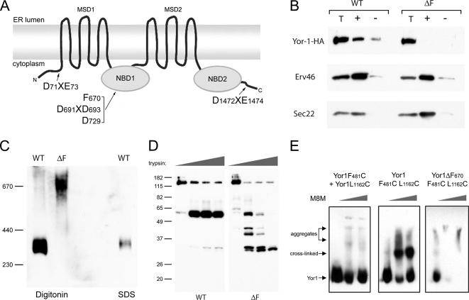
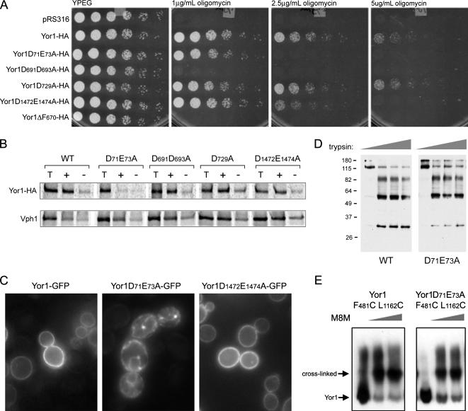
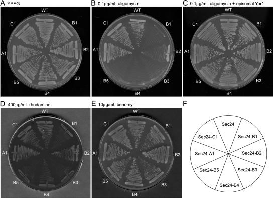
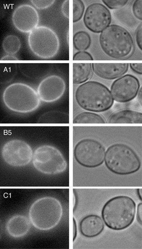
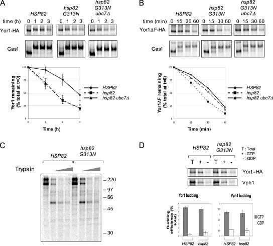
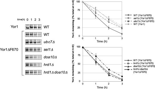
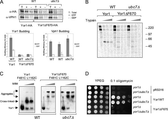
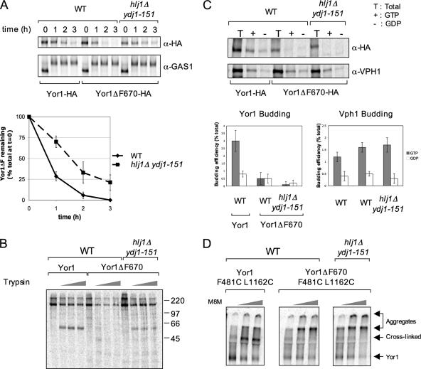
Capture of newly synthesized proteins into endoplasmic reticulum (ER)-derived coat protomer type II (COPII) vesicles represents a critical juncture in the quality control of protein biogenesis within the secretory pathway. The yeast ATP-binding cassette transporter Yor1p is a pleiotropic drug pump that shows homology to the human cystic fibrosis transmembrane conductance regulator (CFTR). Deletion of a phenylalanine residue in Yor1p, equivalent to the major disease-causing mutation in CFTR, causes ER retention and degradation via ER-associated degradation. We have examined the relationship between protein folding, ERAD and forward transport during Yor1p biogenesis. Uptake of Yor1p into COPII vesicles is mediated by an N-terminal diacidic signal that likely interacts with the "B-site" cargo-recognition domain on the COPII subunit, Sec24p. Yor1p-DeltaF is subjected to complex ER quality control involving multiple cytoplasmic chaperones and degradative pathways. Stabilization of Yor1p-DeltaF by inhibiting its degradation does not permit access of Yor1p-DeltaF to COPII vesicles. We propose that the ER quality control checkpoint engages misfolded Yor1p even after it has been stabilized by inhibition of the degradative pathway.


Figures

Figure 1.
Figure 1.
Yor1p-ΔF is excluded from COPII vesicles and is misfolded. (A) Schematic diagram of the yeast ABC transporter, Yor1p, which, like CFTR, has two membrane-spanning domains (MSD1 and MSD2), two nucleotide binding domains (NBD1 and NBD2), and an analogous phenylalanine residue (F670) in NBD1 that is necessary for protein stability and trafficking. Yor1p also has two previously identified terminal diacidic motifs (D71E73 and D1472E1474) and two additional acidic motifs in NBD1 (D691D693 and D729) that may interact with COPII coat machinery in facilitating ER export. (B) Capture of Yor1p into ER-derived COPII vesicles was examined in an in vitro vesicle budding assay by using microsomal membranes expressing HA-tagged forms of Yor1p as the donor membranes. Membranes were incubated with COPII proteins in the presence of GTP (+) or GDP (−), and vesicles were separated from the total membrane (T) fraction by centrifugation. Cargo proteins were detected by immunoblot analysis. (C) Yor1p and Yor1p-ΔF were analyzed by BNGE after solubilization with either digitonin or SDS as indicated followed by immunoblotting to detect HA-tagged forms of Yor1p. The amount of material loaded following solubilization with SDS represents a 1/5 dilution compared with that loaded for digitonin-solubilized samples. (D) Microsomal membranes expressing HA-tagged forms of Yor1p were subjected to limited proteolysis by treating membranes with increasing amounts of trypsin followed by SDS-PAGE and immunoblotting to detect HA-tagged proteolytic fragments. (E) Membranes expressing HA-tagged forms of cysteine-modified Yor1p were cross-linked with increasing concentrations of M8M before SDS-PAGE and immunoblot analysis. Yor1p expressed from two separate plasmids, each bearing single cysteine substitutions showed no cross-linked species (left), whereas Yor1p containing both F481C and L1162C mutations was readily cross-linked into a higher-molecular-weight species (middle). Yor1p-ΔF containing the same substitutions instead presented as a very high-molecular-weight aggregate, with neither the wild-type nor cross-linked species detectable (right).
Figure 2.
Figure 2.
Identification of an N-terminal diacidic motif as the ER exit signal for Yor1p. (A) Four-fold serial dilutions of yeast expressing HA-tagged forms of Yor1p were tested for sensitivity to increasing concentrations of oligomycin (1, 2.5, and 5 μg/ml). Cells expressing wild-type Yor1p were able to grow under all conditions, whereas cells containing the plasmid alone (pRS316) were sensitive to very low levels of oligomycin. The various diacidic mutants showed a range of oligomycin sensitivities, whereas the ER-retained Yor1p-ΔF was completely oligomycin sensitive. (B) Packaging of Yor1p mutants into COPII vesicles was examined by an in vitro vesicle budding assay. Cells were radiolabeled with [35S]methionine/cysteine for 15 min, permeabilized, and incubated with COPII proteins in the presence of either GTP (+) or GDP (−). HA-tagged wild-type and mutant forms of Yor1p were immunoprecipitated and analyzed by SDS-PAGE and PhosphorImage analysis. The percentage of Yor1p found in the vesicle fraction was quantified relative to the total Yor1p (T) present in the donor membranes. Packaging of D71XE73A was severely impaired, whereas that of the D691XD693A, D729A, and D1472XE1474 mutants was similar to that of wild-type Yor1p. An independent cargo protein, Vph1, was also examined to indicate efficient generation of vesicles in the in vitro assay. (C) Wild-type cells coexpressing pRS315–PDR1-3 and Yor1p–GFP fusions as indicated were examined by epifluorescence microscopy. Only the N-terminal di-acidic mutant showed perinuclear staining consistent with ER retention. Wild-type Yor1p and the C-terminal diacidic mutant both localized to the plasma membrane. (D) Folding and stability of Yor1p and the N-terminal diacidic mutant were analyzed by limited proteolysis. Whole cell lysates containing HA-tagged wild-type or mutant forms of Yor1p were incubated with increasing amounts of trypsin, subjected to SDS-PAGE and Yor1p fragments detected by immunoblotting with an anti-HA antibody. (E) The arrangement of transmembrane domains in the N-terminal di-acidic mutant was probed by cysteine cross-linking as described in Figure 1E.
Figure 3.
Figure 3.
Identification of the Sec24p B-site as the cargo capture site for Yor1p. Strains expressing WT Sec24p or various A-, B-, or C-site Sec24p mutants as the sole copy of Sec24p were tested for growth defects in the presence of various compounds as indicated. (A) All strains grew normally on YPEG plates. (B) B-site mutants were unable to grow on plates containing oligomycin whereas A- and C-site mutants grew normally. (C) Oligomycin sensitivity of the B-site mutants could be rescued by transforming these strains with an extra episomal copy of Yor1p. (D) The B-site mutants also showed growth defects in the presence of rhodamine. (E) All strains grew normally in the presence of benomyl.
Figure 4.
Figure 4.
Yor1p-GFP accumulates in the ER in a Sec24-B mutant. Yor1p-GFP was introduced into strains expressing either wild-type Sec24p or A-, B- or C-site Sec24p mutants as indicated. Localization of Yor1p was examined by epifluorescence microscopy (left) and cell integrity monitored by differential interference contrast microscopy (right). In wild-type cells as well as in the A- and C-site mutants, Yor1p-GFP was located at the plasma membrane, whereas in B-site Sec24p mutant cells, the protein showed perinuclear localization consistent with ER-retention in these cells.
Figure 5.
Figure 5.
Yor1p and Yor1p-ΔF are destabilized in a hsp90 mutant strain. (A) Wild-type (HSP82), hsp82ts (hsc82Δ/HSP82G313N), or hsp82ts/ubc7Δ strains expressing an HA-tagged form of Yor1p were grown at 30°C, pulse labeled for 5 min with [35S]methionine/cysteine at 37°C, and chased at 37°C for the times indicated. Cells were subjected to immunoprecipitation for both Yor1p and Gas1p, and the abundance and maturation of these proteins were quantified by SDS-PAGE and PhosphorImage analysis. The results shown are the average of three independent experiments. (B) Wild-type and hsp82 mutants expressing HA-tagged Yor1p-ΔF were analyzed as described in A. (C) The conformation of Yor1p was probed by limited proteolysis. Cells expressing Yor1p were grown at 30°C and pretreated for 25 min at 37°C before pulse labeling with [35S]methionine/cysteine. Cells were permeabilized, and the washed membranes were incubated on ice for 10 min with increasing concentrations of trypsin (0, 100, 200, and 400 ng/μl). Yor1p fragments were immunoprecipitated and visualized by SDS-PAGE and PhosphorImage analysis. (D) Packaging of Yor1p into COPII vesicles was examined by an in vitro vesicle budding assay from cells that had been radiolabeled with [35S]methionine/cysteine at 37°C, permeabilized and incubated with COPII proteins in the presence of either GTP (+) or GDP (−). HA-tagged Yor1p was immunoprecipitated and analyzed by SDS-PAGE and PhosphorImage analysis. The percentage of Yor1p found in the vesicle fraction was quantified relative to the total Yor1p (T) present in the donor membranes. An independent cargo protein, Vph1p, was also examined to indicate efficient generation of vesicles in the in vitro assay.
Figure 6.
Figure 6.
Yor1p-ΔF uses multiple ERAD pathways. HA-tagged Yor1p or Yor1p-ΔF was expressed in either a wild-type strain or strains deleted for a variety of ERAD machineries as indicated. Cells were pulse-labeled for 5 min with [35S]methionine/cysteine and chased for the times indicated. Yor1p was immunoprecipitated from cell lysates and analyzed by SDS-PAGE and PhosphorImage analysis. The results shown are the average of three independent experiments.
Figure 7.
Figure 7.
Biogenesis and trafficking of Yor1p-ΔF in a ubc7Δ mutant strain. (A) Permeabilized cells from wild type and ubc7Δ strains expressing HA-tagged Yor1p or Yor1p-ΔF were used in an in vitro vesicle budding assay. Total membranes (T) and budded vesicles were collected from reactions that contained either GTP (+) or GDP (−). Yor1p and Vph1 packaging into vesicles was quantified by immunoprecipitation followed by SDS-PAGE and PhosphorImage analysis. (B) Radiolabeled permeabilized cells from a wild-type strain and a ubc7Δ strain containing HA-tagged Yor1p or Yor1p-ΔF were incubated on ice for 10 min with increasing concentrations of trypsin (0, 100, 200, and 400 ng/μl). Yor1p fragments were immunoprecipitated from cell lysates and analyzed by SDS-PAGE and PhosphorImage analysis. (C) Whole cell lysates from wild-type or ubc7Δ strains expressing cysteine-substituted forms of Yor1p or Yor1p-ΔF were cross-linked with increasing concentrations of M8M as described in Figure 1E. Stabilization of Yor1p-ΔF in the ubc7Δ strain did not result in the appearance of the wild-type cross-linked species. (D) Yor1p function at the plasma membrane was tested phenotypically by assessing growth in the presence of oligomycin. Strains bearing deletions in YOR1 or YOR1 and UBC7 were transformed with the plasmids indicated and serial dilutions spotted onto YPEG or YPEG supplemented with oligomycin.
Figure 8.
Figure 8.
Yor1p-ΔF is stabilized in a hsp40 mutant strain. (A) Wild-type and hsp40 (hlj1Δ/ydj1-151) strains expressing HA-tagged Yor1p or Yor1p-ΔF were grown at 30°C and pretreated for 25 min at 37°C. Then, strains were pulse-labeled for 5 min with [35S]methionine/cysteine at 37°C and chased at 37°C for the times indicated. Yor1p and Gas1p were immunoprecipitated from cell lysates and analyzed by SDS-PAGE and PhosphorImage analysis. The results shown are the average of three independent experiments. (B) Permeabilized cells from wild-type and hsp40 (hlj1Δ/ydj1-151) strains that had been pretreated at 37°C before labeling were used in an in vitro vesicle budding assay. Total membranes (T) and budded vesicles were collected from reactions that contained either GTP (+) or GDP (−). Yor1p and Vph1 packaging into vesicles was quantified by immunoprecipitation followed by SDS-PAGE and PhosphorImage analysis. (C) Radiolabeled permeabilized cells from wild type and hsp40 (hlj1Δ/ydj1-151) strains expressing HA-tagged Yor1p or Yor1p-ΔF at restrictive temperature were incubated on ice for 10 min with increasing concentrations of trypsin (0, 100, 200, and 400 ng/μl). Fragments were immunoprecipitated from cell lysates and analyzed by SDS-PAGE and PhosphorImage analysis. (D) Radiolabeled permeabilized cells from wild-type and hsp40 (hlj1Δ/ydj1-151) strains expressing cysteine-substituted forms of Yor1p or Yor1p-ΔF at restrictive temperature were subjected to cross-linking with M8M before solublization and immunoprecipitation. Stabilization of Yor1p-ΔF in the hsp40 mutant did not result in the appearance of the typical cross-linked species.

Similar articles

Cited by

References

    1. Ahner A., Brodsky J. L. Checkpoints in ER-associated degradation: excuse me, which way to the proteasome? Trends Cell Biol. 2004;14:474–478. - PubMed
    1. Barlowe C., Orci L., Yeung T., Hosobuchi M., Hamamoto S., Salama N., Rexach M. F., Ravazzola M., Amherdt M., Schekman R. COPII: a membrane coat formed by Sec proteins that drive vesicle budding from the endoplasmic reticulum. Cell. 1994;77:895–907. - PubMed
    1. Bohen S. P., Yamamoto K. R. Isolation of Hsp90 mutants by screening for decreased steroid receptor function. Proc. Natl. Acad. Sci. USA. 1993;90:11424–11428. - PMC - PubMed
    1. Borkovich K. A., Farrelly F. W., Finkelstein D. B., Taulien J., Lindquist S. hsp82 is an essential protein that is required in higher concentrations for growth of cells at higher temperatures. Mol. Cell. Biol. 1989;9:3919–3930. - PMC - PubMed
    1. Bukau B., Weissman J., Horwich A. Molecular chaperones and protein quality control. Cell. 2006;125:443–451. - PubMed

Publication types

MeSH terms