. 2007 Aug 17;101(4):420-8.
doi: 10.1161/CIRCRESAHA.107.156463. Epub 2007 Jul 5.

Abl silencing inhibits CAS-mediated process and constriction in resistance arteries

Affiliations

Abl silencing inhibits CAS-mediated process and constriction in resistance arteries

Yana Anfinogenova et al. Circ Res. .

Abstract

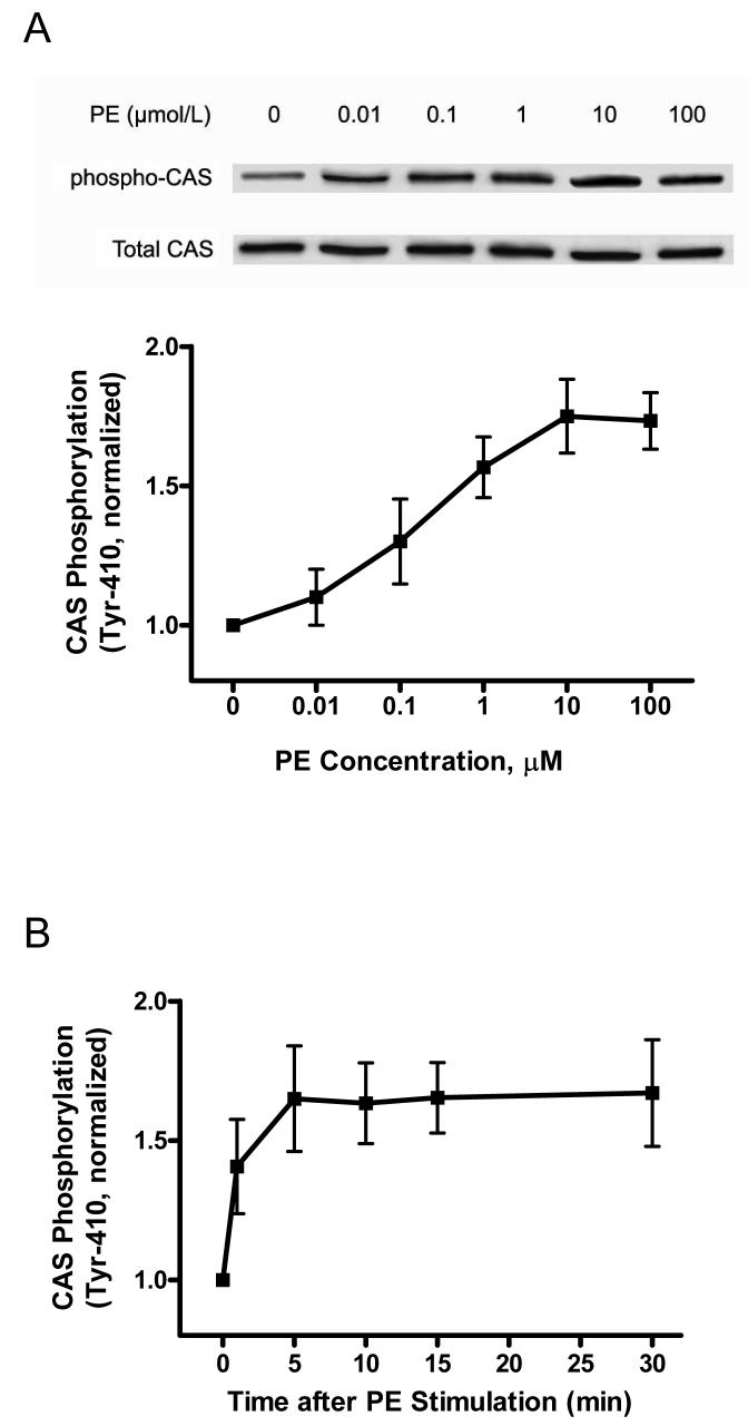
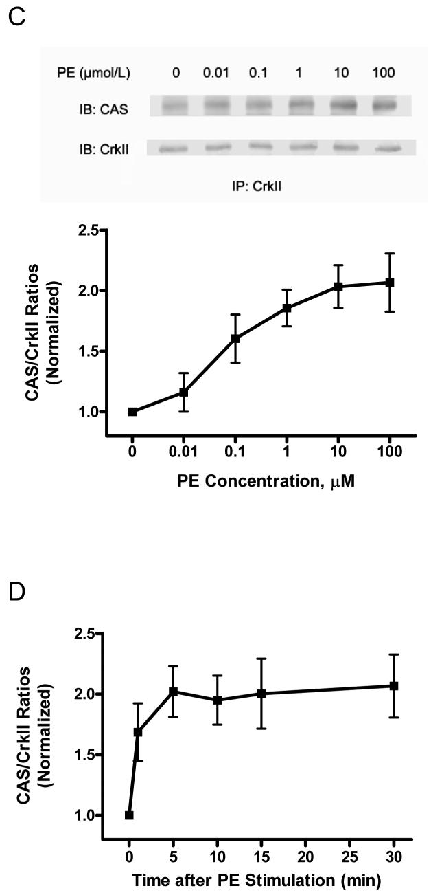
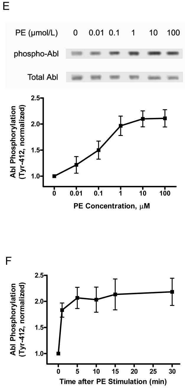
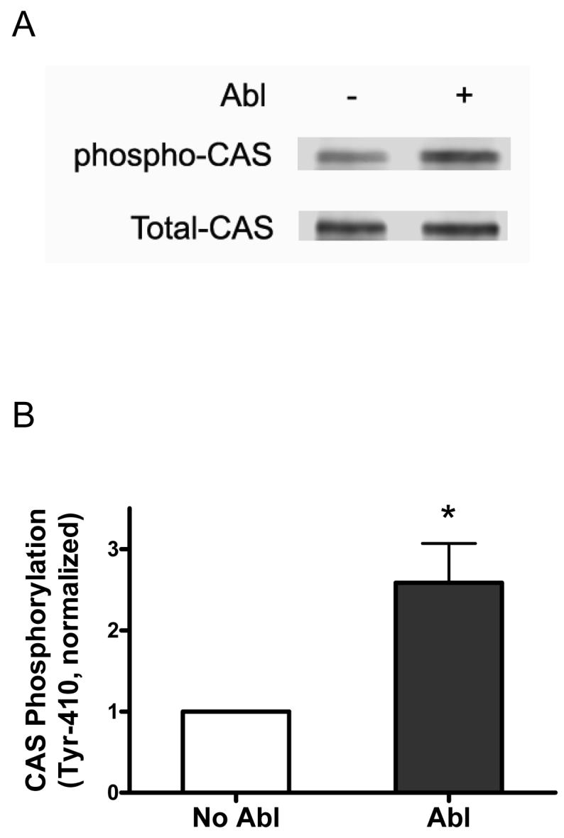
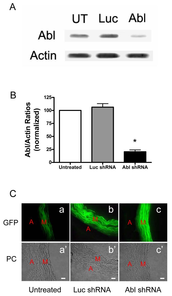
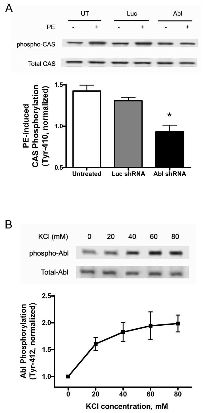
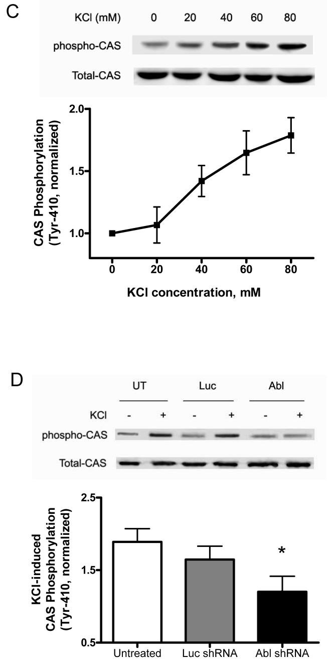
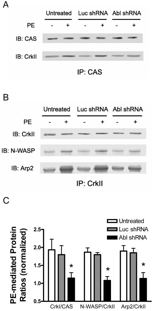
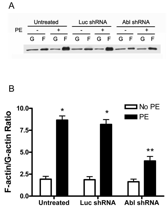
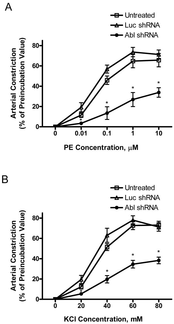
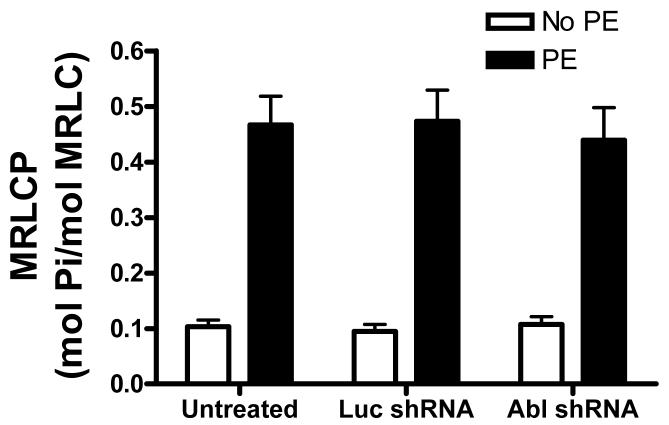
The tyrosine phosphorylated protein Crk-associated substrate (CAS) has previously been shown to participate in the cellular processes regulating dynamic changes in the actin architecture and arterial constriction. In the present study, treatment of rat mesenteric arteries with phenylephrine (PE) led to the increase in CAS tyrosine phosphorylation and the association of CAS with the adapter protein CrkII. CAS phosphorylation was catalyzed by Abl in an in vitro study. To determine the role of Abl tyrosine kinase in arterial vessels, plasmids encoding Abl short hairpin RNA (shRNA) were transduced into mesenteric arteries by chemical loading plus liposomes. Abl silencing diminished increases in CAS phosphorylation on PE stimulation. Previous studies have shown that assembly of the multiprotein compound containing CrkII, neuronal Wiskott-Aldrich Syndrome Protein (N-WASP) and the Arp2/3 (Actin Related Protein) complex triggers actin polymerization in smooth muscle as well as in nonmuscle cells. In this study, Abl silencing attenuated the assembly of the multiprotein compound in resistance arteries on contractile stimulation. Furthermore, the increase in F/G-actin ratios (an index of actin assembly) and constriction on contractile stimulation were reduced in Abl-deficient arterial segments compared with control arteries. However, myosin regulatory light chain phosphorylation (MRLCP) elicited by contractile activation was not inhibited in Abl-deficient arteries. These results suggest that Abl may play a pivotal role in mediating CAS phosphorylation, the assembly of the multiprotein complex, actin assembly, and constriction in resistance arteries. Abl does not participate in the regulation of myosin activation in arterial vessels during contractile stimulation.


Figures

Figure 1
Figure 1. Activation with phenylephrine (PE) results in increases in CAS phosphorylation, the association of CAS with CrkII and Abl phosphorylation in resistance arteries
Unstimulated or PE-stimulated rat mesenteric arteries were frozen for biochemical analysis. (A) Representative blots show the dose-dependent enhancement of CAS phosphorylation (Tyr-410) upon PE stimulation (5 min). The level of CAS phosphorylation elicited by PE is normalized to the value of unstimulated arteries (n = 4-5). (B) CAS phosphorylation upon PE stimulation (10 μmol/L) is time-dependent (n = 4). (C) Representative blots illustrate the increased amount of CAS co-immunoprecipitated with CrkII upon PE stimulation (5 min). CAS/CrkII ratios stimulated by PE are expressed as magnitude of the level of unstimulated arteries (n = 4). (D) The time-dependent increase in CAS/CrkII coupling in response to PE (10 μmol/L) stimulation (n = 4-5). (E) Representative blots illustrate the effects of PE stimulation (5 min) on Abl phosphorylation (Tyr-412). PE-stimulated Abl phosphorylation is normalized to the level of unstimulated tissues (n = 4-6). (F) The time-dependent enhancement of Abl phosphorylation upon PE (10 μmol/L) stimulation (n = 4). Values represent mean ± SE.
Figure 2
Figure 2. Abl catalyzes CAS phosphorylation in vitro
(A) The immunoblots illustrate the phosphorylation of CAS (Tyr-410) by Abl. Abl-mediated CAS phosphorylation 30 min after the initiation of reaction was determined as described under “Materials and Methods.” (B) The phosphorylation level after Abl treatment (30 min) is normalized to the level of CAS phosphorylation in the absence of Abl. *Significantly higher phosphorylation levels in the presence of Abl compared to the level without Abl treatment (n = 3, P < 0.05).
Figure 3
Figure 3. Silencing of Abl gene in mesenteric segments by short hairpin RNA (shRNA)
(A) Blots of protein extracts from untreated arteries (UT) or arteries that had been cultured for 2 days with plasmids encoding luciferase shRNA (Luc) or Abl shRNA (Abl) were probed with antibodies against Abl and α-actin. The level of Abl was lower in arteries producing Abl shRNA than in untreated arteries or segments generating Luc shRNA. Similar amounts of actin were detected in all three samples. (B) Abl/actin ratios in arteries producing Luc shRNA or Abl shRNA are normalized to ratios obtained in tissues not treated with plasmids (n = 5). Values are mean ± SE. *Significantly lower protein ratios in segments producing Abl shRNA compared to untreated arteries or arteries generating Luc shRNA (P < 0.01). (C) Representative images showing green fluorescence protein (GFP) signals observed in the medial smooth muscle layer of the arterial walls treated with plasmids, indicating effective transfection in the smooth muscle layer of arteries. Weak autofluorescence (panel a) was observed from the untreated mesenteric artery (panel a'). GFP signal (panel b) was detected from the artery transfected with plasmids encoding luciferase (Luc) shRNA (panel b'); the fluorescence (panel c), the artery transfected with plasmids for Abl shRNA (panel c'). PC, phase contrast images. A, adventitia; M, media. Intima was removed during preparation. Bar, 15 μm.
Figure 4
Figure 4. CAS phosphorylation in response to contractile stimulation is diminished in Abl-deficient arteries
(A) Blots of extracts from resistance arteries that had been treated with plasmids encoding Luc shRNA or Abl shRNA, or untreated arteries were detected with use of phospho-CAS (Tyr-410) antibody, stripped, and reprobed with (total) CAS antibody. CAS phosphorylation upon stimulation with PE was depressed in Abl-deficient arteries (n = 5). Values are normalized to the phosphorylation level in corresponding unstimulated arteries. (B) Representative blots illustrate the effects of KCl stimulation (5 min) on Abl phosphorylation (Tyr-412). KCl-stimulated Abl phosphorylation is normalized to the level of unstimulated tissues (n = 5). (C) Representative blots show the increase in CAS phosphorylation (Tyr-410) upon KCl stimulation (5 min). The level of CAS phosphorylation elicited by KCl is normalized to the value of unstimulated arteries (n = 5). (D) Representative blots illustrate the effects of Abl silencing on KCl-induced CAS phosphorylation. Values are normalized to the phosphorylation level in corresponding unstimulated arteries (n = 5). Values are mean ± SE. *Significantly lower levels upon PE or KCl stimulation compared to corresponding untreated (control) arteries or tissues treated with Luc shRNA (P < 0.01).
Figure 5
Figure 5. Association of CAS with CrkII and formation of protein complex containing CrkII are attenuated in Abl-deficient arteries upon PE stimulation
Untreated arteries or arteries that had been treated with plasmids encoding Luc shRNA, or Abl shRNA were stimulated with PE, or left unstimulated. (A) Blots of CAS immunoprecipitates from these arteries were probed with antibodies against CAS and CrkII. The immunoblots illustrate the effects of Abl silencing on CAS/CrkII coupling. (B) Blots of CrkII immunoprecipitates from these arteries were probed with antibodies against CrkII, N-WASP, and Arp2. Abl silencing inhibits the complex formation. (C) Individual protein ratios are normalized to protein ratios in corresponding unstimulated arteries. *Significantly lower protein ratios in response to PE stimulation compared to corresponding PE-induced protein ratios in untreated arteries or arteries producing Luc shRNA (n = 3, P < 0.05). Values are mean ± SE.
Figure 6
Figure 6. Abl silencing inhibits the increase in F/G-actin ratios upon PE activation
Arteries producing Luc shRNA or Abl shRNA were stimulated with PE, or left unstimulated. F/G-actin ratios were then assessed. (A) The representative blot illustrating the effects of Abl silencing on the amounts of G-actin and F-actin. (B) *Significantly higher F/G-actin ratios upon PE stimulation compared to corresponding unstimulated values (P < 0.01). **PE-stimulated F/G-actin ratios in tissues producing Abl shRNA are significantly lower than those in untreated tissues or arteries generating Luc shRNA (P < 0.05). Values are mean ± SE (n = 5).
Figure 7
Figure 7. Constriction is diminished in Abl-deficient arteries in response to activation with PE or KCl
(A) Compared to untreated tissues or arteries producing luciferase shRNA (Luc RNA), PE-induced dose-response curve in arteries producing Abl shRNA was right shifted. Maximal contraction was also significantly reduced (n = 4-6). (B) The dose-response curve in response to KCl stimulation was right shifted with the reduced maximal response compared to untreated tissues or arteries producing Luc RNA. Constriction in response to PE or KCl is normalized to corresponding maximal levels in each artery before incubation (n = 5). Values are mean ± SE. Asterisk indicates significantly lower response as compared to untreated tissues or tissues generating Luc shRNA (P < 0.05).
Figure 8
Figure 8. Effects of Abl silencing on myosin regulatory light chain phosphorylation (MRLCP)
Increases in MRLCP upon PE stimulation are similar in arteries that have been treated with plasmids encoding Luc shRNA or Abl shRNA, or no plasmids (P > 0.05). Values shown are mean ± SE (n = 5).

References

    1. Tang DD, Zhang W, Gunst SJ. The Adapter Protein CrkII Regulates Neuronal Wiskott-Aldrich Syndrome Protein, Actin Polymerization, and Tension Development during Contractile Stimulation of Smooth Muscle. J Biol Chem. 2005 June 17;280(24):23380–9. - PubMed
    1. Tang DD, Gunst SJ. The small GTPase Cdc42 regulates actin polymerization and tension development during contractile stimulation of smooth muscle. J Biol Chem. 2004 December 10;279(50):51722–8. - PubMed
    1. Zhang W, Wu Y, Du L, Tang DD, Gunst SJ. Activation of the Arp2/3 complex by N-WASp is required for actin polymerization and contraction in smooth muscle. Am J Physiol Cell Physiol. 2005 May;288(5):C1145–C1160. - PubMed
    1. Barany M, Barron JT, Gu L, Barany K. Exchange of the actin-bound nucleotide in intact arterial smooth muscle. J Biol Chem. 2001 December 21;276(51):48398–403. - PubMed
    1. Cipolla MJ, Gokina NI, Osol G. Pressure-induced actin polymerization in vascular smooth muscle as a mechanism underlying myogenic behavior. FASEB J. 2002 January;16(1):72–6. - PubMed

Publication types

MeSH terms