. 2007 Jun 8:13:823-32.

Human embryonic and neuronal stem cell markers in retinoblastoma

Affiliations

Human embryonic and neuronal stem cell markers in retinoblastoma

Gail M Seigel et al. Mol Vis. .

Abstract

Purpose: Retinoblastoma (RB) is the most common intraocular tumor of early childhood. The early onset of RB, coupled with our previous findings of cancer stem cell characteristics in RB, led us to hypothesize that subpopulations of RB tumors harbor markers and behaviors characteristic of embryonic and neuronal origin.

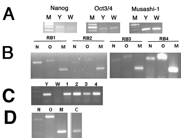
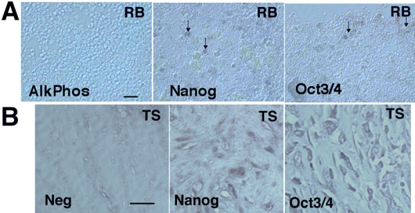
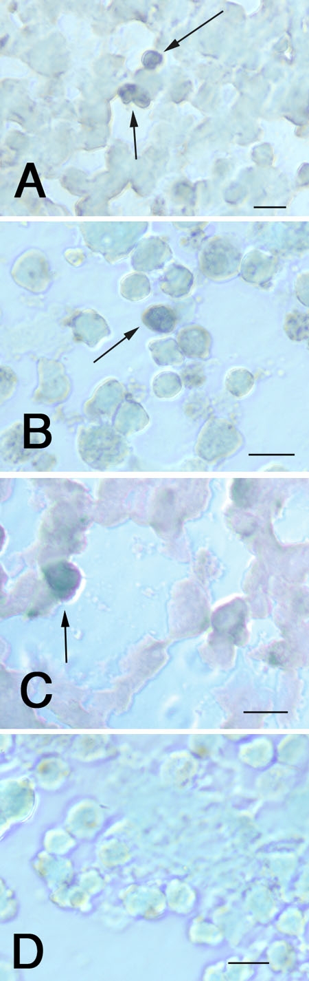
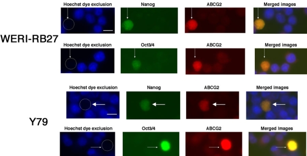
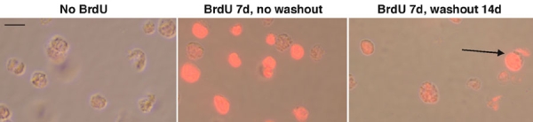
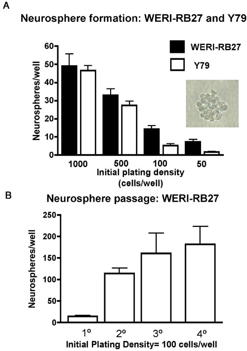
Methods: Our RB sources included: human pathological tissues, and the human RB cell lines Y79 and WERI-RB27. Microarray screening, single and dual-label immunocytochemistry and RT-PCR were performed to detect embryonic and neuronal stem cell markers, such as Oct3/4, Nanog, CD133, and Musashi-1. To test for functional evidence of stem cell behavior, we examined RB cells for their ability to form neurospheres and retain BrdU label as indicators of self-renewal and slow cell cycling, respectively.

Results: Microarray comparisons of human RB tumors with normal retinal tissue detected upregulation of a number of genes involved in embryonic development that were also present in Y79 cells, including Oct3/4, Nanog, Musashi-1 and Musashi-2, prominin-1 (CD133), Jagged-2, Reelin, Thy-1, nestin, Meis-1,NCAM, Patched, and Notch4. Expression of Musashi-1, Oct3/4 and Nanog was confirmed by immunostaining and RT-PCR analyses of RB tumors and RB cell lines. CD133 expression was confirmed by PCR analysis. Y79 and WERI-RB27 contained populations of Hoechst-dim/ABCG2-positive cells that co-localized with embryonic stem cell markers Oct3/4-ABCG2 and Nanog-ABCG2. Subpopulations of Y79 and WERI-RB27 cells were label-retaining (as seen by BrdU incorporation) and were able to generate neurospheres, both hallmarks of a stem cell phenotype.

Conclusions: Small subpopulation(s) of RB cells express human embryonic and neuronal stem cell markers. There are also subpopulations that demonstrate functional behavior (label retention and self-renewal) consistent with cancer stem cells. These findings support the hypothesis that RB is a heterogeneous tumor comprised of subpopulation(s) with stem cell-like properties.


Figures

Figure 1
Figure 1
Detection of Oct 3/4, Nanog Musashi-1, and CD133 in human retinoblastoma tumors, retinoblastoma cell lines and normal human retina by reverse transcriptase polymerase chain reaction. Human retinoblastoma (RB) tumors and cell lines were examined by RT-PCR as described in Methods. A: Nanog, Oct3/4, and Musashi gene expression in WERI-RB27 and Y79 cell lines. M represents marker; Y represents Y79; W represents WERI-RB27. B: Nanog (N), Oct3/4 (O), and Musashi-1 (M) expression in four human RB tumors (RB1, 2, 3, 4). C: CD133 expression seen in Y79 cells (Y) and RB tumors (1,2,3,4), but not WERI-RB27 cells (W). D: Nanog (N), Oct3/4 (O), Musashi-1 (M) and CD133 (C) expression in normal human retina.
Figure 2
Figure 2
Human retinoblastoma tumors exhibit immunoreactivity to human embryonic stem cell markers Oct 3/4 and Nanog. A: Human retinoblastoma (RB) tumors were prepared as frozen sections, and immunostained for human embryonic stem cell markers AlkPhos, Oct3/4, and Nanog, Immunoreactivity to Oct3/4 and Nanog was evident (eg. arrows), while no reaction was seen AlkPhos. B: Human testicular seminoma tissue (TS) was used as a positive control for Oct3/4 and Nanog. The scale bars represents 5 μm.
Figure 3
Figure 3
Musashi-1 in retinoblastoma tumors and cell lines. Human retinoblastoma (RB) tumors and cell lines were prepared as frozen sections, and immunostained for Musashi-1: A: Human RB tumor; B: Y79 cells; C: WERI-RB27 cells; D: Negative control. The scale bar represents 5 μm. Arrows indicate positive cells.
Figure 4
Figure 4
Colocalization of Nanog or Oct 3/4 in Hoechst-dim/ABCG2 positive cells. Y79 and WERI-RB27 human retinoblastoma cells were examined for fluorescent Hoechst 33342 dye uptake, ABCG2 immunoreactivity, coupled with either Nanog or Oct3/4 immunoreactivity. Each horizontal panel depicts the same microscopic field, viewed under separate fluorescent filters for Hoechst, FITC and TRITC, as well as a merged image of all three fields. As seen in the "Hoechst dye exclusion" field, the arrow points to a cell that has excluded the Hoechst dye and appears "Hoechst dim". This is due to the active Hoechst dye exclusion properties of the ABCG2 protein. In the next two panels, we see the same cell, as indicated by the arrow, that is immunoreactive for Nanog or Oct3/4 and ABCG2. When the three images are merged, ABCG2 colocalizes with both Nanog and Oct 3/4. The scale bar represents 5 μm.
Figure 5
Figure 5
Label-retaining cells in retinoblastoma cultures. Y79 human retinoblastoma cells were pulsed with 10 μM BrdU for 4-7 days and washed out for 14 days. At 14 days, BrdU-immunoreactive cells comprised approximately 3-4% of the population. A: No BrdU added (negative control); B: BrdU added for 7 days without washout. C: BrdU for 7 days and 14 days of washout. Three percent of cells were still labeled after 14 days (arrow). The scale bar represents 5 μm.
Figure 6
Figure 6
Neurosphere formation in retinoblastoma cultures. A: Y79 and WERI-R27 cells were plated as single cell suspensions in 96 well dishes at initial plating densities of 50-1,000 cells per well, in triplicate. Low cell densities were chosen to minimize effects of non-specific cell aggregation in favor of neurospheres originating from one single cell. After five days, neurospheres were counted and results presented. Both Y79 and WERI-RB27 human retinoblastoma cells formed neurospheres at all cell densities tested. A typical neurosphere (WERI-RB27) is shown (inset). B: WERI-RB27 cells were prepared as single cell suspensions at an initial plating density of 100 cells per well, in triplicate. Every 3-4 days, neurospheres were counted and then dissociated into single cell suspensions, diluted 1:2, and replated. The graph depicts the number of neurospheres counted at each passage.

Similar articles

Cited by

References

    1. Gallie BL, Dunn JM, Chan HS, Hamel PA, Phillips RA. The genetics of retinoblastoma. Relevance to the patient. Pediatr Clin North Am. 1991;38:299–315. - PubMed
    1. Abramson DH, Melson MR, Dunkel IJ, Frank CM. Third (fourth and fifth) nonocular tumors in survivors of retinoblastoma. Ophthalmology. 2001;108:1868–76. - PubMed
    1. Wong FL, Boice JD, Jr, Abramson DH, Tarone RE, Kleinerman RA, Stovall M, Goldman MB, Seddon JM, Tarbell N, Fraumeni JF, Jr, Li FP. Cancer incidence after retinoblastoma. Radiation dose and sarcoma risk. JAMA. 1997;278:1262–7. - PubMed
    1. Pardal R, Clarke MF, Morrison SJ. Applying the principles of stem-cell biology to cancer. Nat Rev Cancer. 2003;3:895–902. - PubMed
    1. Reya T, Morrison SJ, Clarke MF, Weissman IL. Stem cells, cancer, and cancer stem cells. Nature. 2001;414:105–11. - PubMed

Publication types

MeSH terms

LinkOut - more resources