

Effects of beta-adrenergic receptor antagonists on oxidative stress in purified rat retinal ganglion cells
- PMID: 17615544
- PMCID: PMC2770200
Effects of beta-adrenergic receptor antagonists on oxidative stress in purified rat retinal ganglion cells
Abstract
Purpose: To investigate the effect of beta-adrenergic receptor antagonists against oxidative stress on purified rat retinal ganglion cells (RGCs), timolol, betaxolol, carteolol and nipradilol were included in the present study.
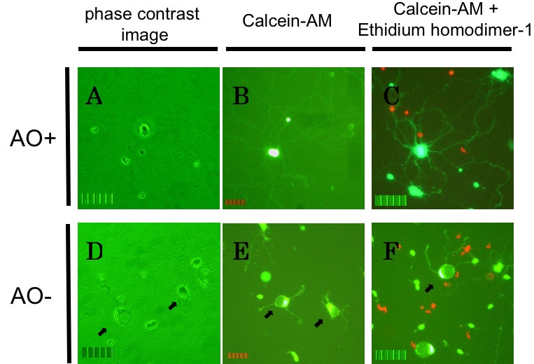
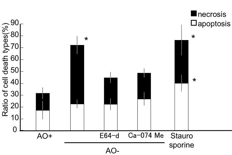
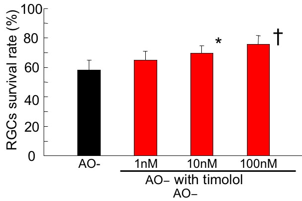
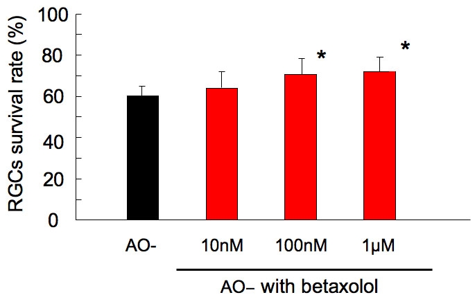
Methods: RGCs were purified using a 2 step panning procedure from postnatal days 6-8 using Wistar rats. After 72 h in culture under normal condition, RGCs were exposed to oxidative stress induced by B27 medium without anti-oxidant. To verify whether this stress is apoptotic or necrotic, Annexin V and propidium iodide were used to detect apoptotic and necrotic cells after 2 h stress. The presence of a proinhibitor for intracellular cathepsin B, and an inhibitor for thiol protease (cathepsin B/H/L, calpain), was also assessed to verify necrotic cell death event in oxidative conditions. Next, RGC cultures under oxidative stress were incubated with timolol, betaxolol, carteolol, and nipradilol added, respectively, for 24 h culture. The RGC viability in each condition normalized to that under normal condition was evaluated as live cell percentage based on total experiments of 8-15.
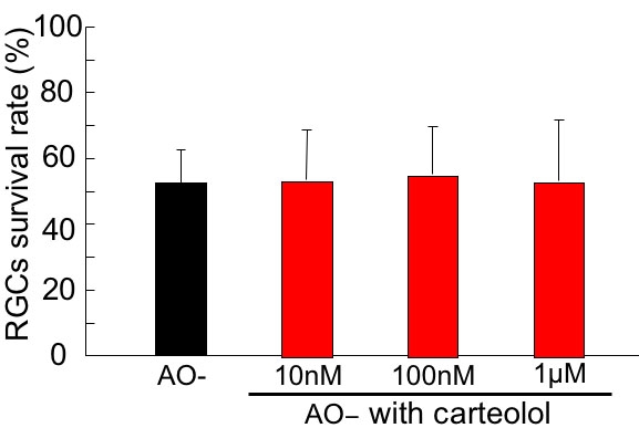
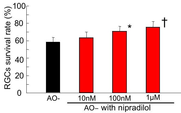
Results: Two h after oxidative stress, Annexin V and propidium iodide positive cells increased. Increased cell death under oxidative stress was significantly reduced by inhibitors for cathepsin or calpain. These data suggest that increased cell death under the current oxidative stress was due to necrosis. Under oxidative stress for 24 h, RGC viability reduced to 52.5-60.2% as compared with normal. With 10 nM and 100 nM timolol, live cell significantly increased to 69.3% and 75.5%, respectively. Both betaxolol and nipradilol enhanced live RGCs significantly in concentration of 100 nM and 1 microM, with viability of 70.5%, 71.6%, and 70.4%, 74.7%, respectively. While with 10 nM, 100 nM and 1 microM addition of carteolol, there was no significant increase in live RGC percentage which ranged from 53.1-55.0%.
Conclusions: Timolol, betaxolol and nipradilol, but not carteolol, showed neuroprotective effects against oxidative stress induced by B27 without antioxidant on purified rat RGCs at concentrations of 10 nM or higher. Although the neuroprotective mechanism of beta-blockers for oxidative stress is still unknown, this additive effect may deserve future studies.
Figures
Similar articles
-
Neuroprotective effect of astaxanthin against rat retinal ganglion cell death under various stresses that induce apoptosis and necrosis.Mol Vis. 2014 Dec 31;20:1796-805. eCollection 2014. Mol Vis. 2014. PMID: 25593507 Free PMC article.
-
Hypoxia-induced retinal ganglion cell death and the neuroprotective effects of beta-adrenergic antagonists.Brain Res. 2007 May 7;1148:28-37. doi: 10.1016/j.brainres.2007.02.027. Epub 2007 Feb 22. Brain Res. 2007. PMID: 17368577
-
Neuroprotective effects of prostaglandin analogues on retinal ganglion cell death independent of intraocular pressure reduction.Exp Eye Res. 2011 Sep;93(3):265-70. doi: 10.1016/j.exer.2011.06.022. Epub 2011 Jul 26. Exp Eye Res. 2011. PMID: 21791206
-
Comparison of the neuroprotective effects of adrenoceptor drugs in retinal cell culture and intact retina.Invest Ophthalmol Vis Sci. 2002 Aug;43(8):2666-76. Invest Ophthalmol Vis Sci. 2002. PMID: 12147601
-
Retinal-cell-conditioned medium prevents TNF-alpha-induced apoptosis of purified ganglion cells.Invest Ophthalmol Vis Sci. 2005 Aug;46(8):2983-91. doi: 10.1167/iovs.04-1177. Invest Ophthalmol Vis Sci. 2005. PMID: 16043875
Cited by
-
Visual field loss in patients with normal-tension glaucoma under topical nipradilol or timolol: subgroup and subfield analyses of the nipradilol-timolol study.Jpn J Ophthalmol. 2010 Jul;54(4):278-85. doi: 10.1007/s10384-010-0815-z. Epub 2010 Aug 11. Jpn J Ophthalmol. 2010. PMID: 20700793 Clinical Trial.
-
TRPV1: contribution to retinal ganglion cell apoptosis and increased intracellular Ca2+ with exposure to hydrostatic pressure.Invest Ophthalmol Vis Sci. 2009 Feb;50(2):717-28. doi: 10.1167/iovs.08-2321. Epub 2008 Oct 24. Invest Ophthalmol Vis Sci. 2009. PMID: 18952924 Free PMC article.
-
Neuroprotective effect of astaxanthin against rat retinal ganglion cell death under various stresses that induce apoptosis and necrosis.Mol Vis. 2014 Dec 31;20:1796-805. eCollection 2014. Mol Vis. 2014. PMID: 25593507 Free PMC article.
-
Neuroprotective effects of flavonoids on hypoxia-, glutamate-, and oxidative stress-induced retinal ganglion cell death.Mol Vis. 2011;17:1784-93. Epub 2011 Jul 2. Mol Vis. 2011. PMID: 21753864 Free PMC article.
-
Carteolol hydrochloride suppresses the generation of reactive oxygen species and rescues cell death after ultraviolet irradiation of cultured lens epithelial cells.Open Ophthalmol J. 2010 Oct 12;4:60-5. doi: 10.2174/1874364101004010060. Open Ophthalmol J. 2010. PMID: 21283534 Free PMC article.
References
-
- Castagne V, Lefevre K, Natero R, Clarke PG, Bedker DA. An optimal redox status for the survival of axotomized ganglion cells in the developing retina. Neuroscience. 1999;93:313–20. - PubMed
-
- Orrenius S, Burkitt MJ, Kass GE, Dypbukt JM, Nicotera P. Calcium ions and oxidative cell injury. Ann Neurol. 1992;32:S33–42. - PubMed
-
- Choi DW. Calcium-mediated neurotoxicity: relationship to specific channel types and role in ischemic damage. Trends Neurosci. 1988;11:465–9. - PubMed
-
- Winkler BS, Boulton ME, Gottsch JD, Sternberg P. Oxidative damage and age-related macular degeneration. Mol Vis. 1999;5:32. http://www.molvis.org/molvis/v5/a32/ - PMC - PubMed
-
- Organisciak DT, Darrow RM, Barsalou L, Darrow RA, Kutty RK, Kutty G, Wiggert B. Light history and age-related changes in retinal light damage. Invest Ophthalmol Vis Sci. 1998;39:1107–16. - PubMed
Publication types
MeSH terms
Substances
LinkOut - more resources
Full Text Sources