. 2007 Jun 12:13:840-53.

Inhibition of NAD(P)H oxidase reduces apoptosis and avascular retina in an animal model of retinopathy of prematurity

Affiliations

Inhibition of NAD(P)H oxidase reduces apoptosis and avascular retina in an animal model of retinopathy of prematurity

Yuta Saito et al. Mol Vis. .

Abstract

Purpose: To study the mechanisms of action of the antioxidants, n-acetylcysteine (NAC), and the nicotinamide adenine dinucleotide phosphate (NAPDH) oxidase oxidase inhibitor, apocynin, on intravitreous neovascularization (IVNV), and retinal avascularity in a rat model of retinopathy of prematurity (ROP).

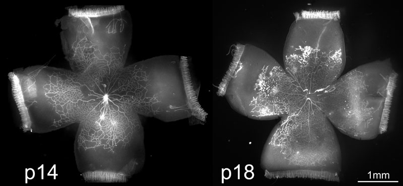
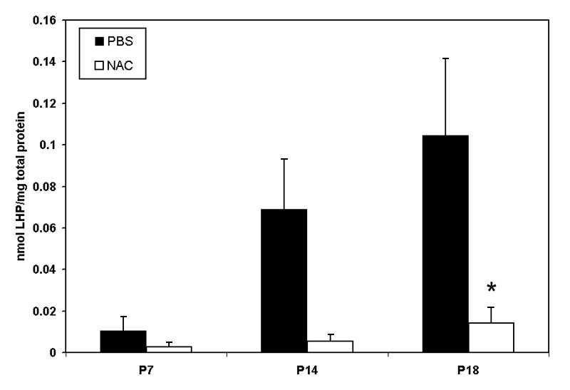
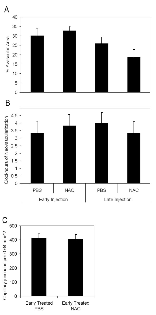
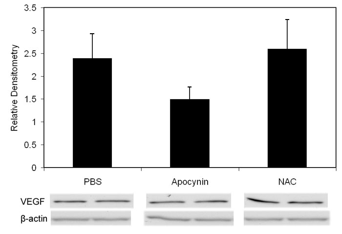
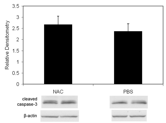
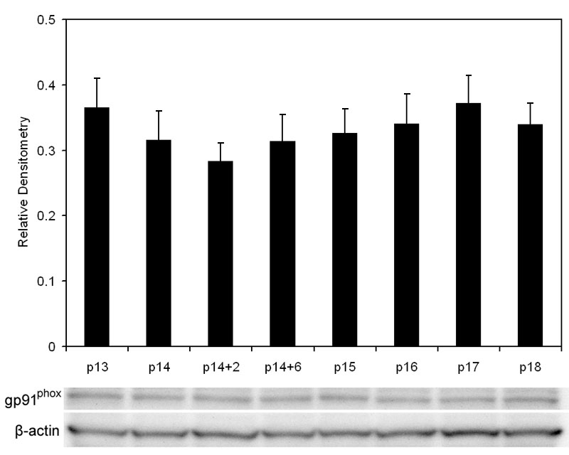
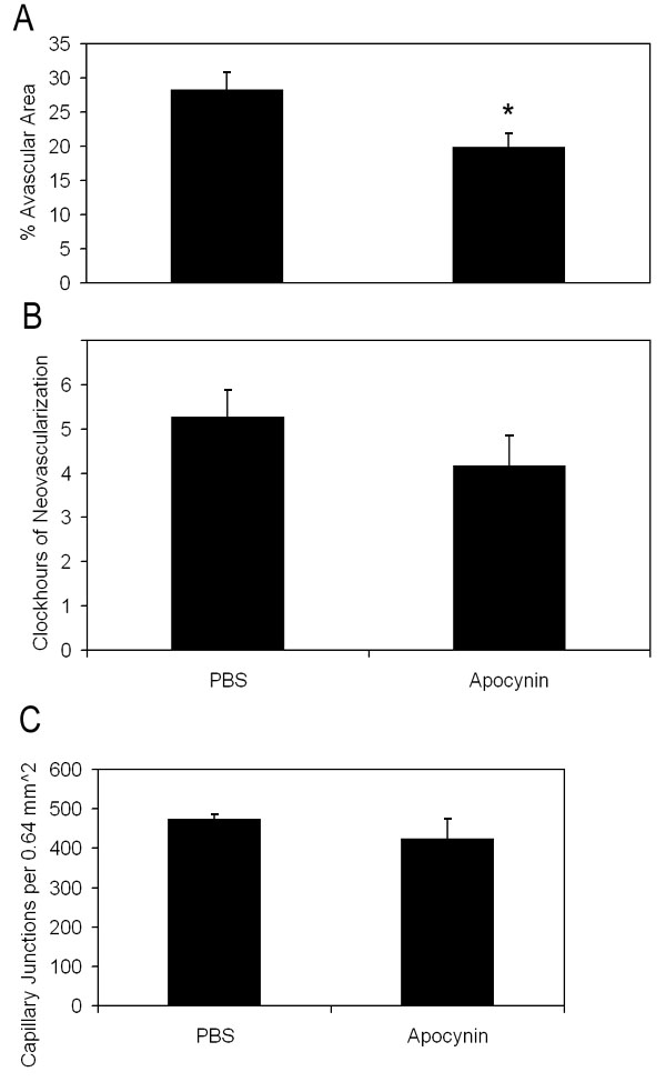
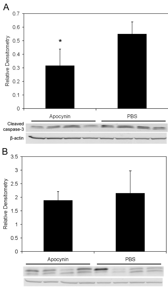
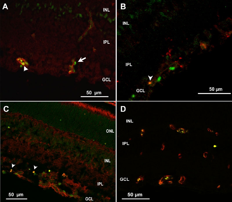
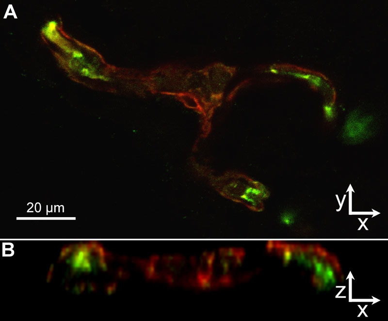
Methods: Newborn rats exposed to oxygen-induced retinopathy underwent intraperitoneal (IP) injections of NAC (150 mg/kg) at post-natal day (p)2, p6, p10 (early NAC-treated), or p12 through p17 (late NAC-treated), apocynin (10 mg/kg) from p12 through p17, or phosphate buffered saline (PBS; controls). Lipid hydroperoxide (LHP) was measured in early NAC-treated oxygen-induced retinopathy (OIR) at p7, p14 and p18. Pups were placed in room air at p14. At p18, retinal flat mounts were scored for IVNV and avascular/total retinal area, or retinas were assayed for cleaved caspase-3 and vascular endothelial growth factor (VEGF) protein. In non-injected OIR pups, retinas were assayed for gp91(phox). Cryosections were stained with isolectin B4, cleaved caspase-3, CD68, CD31, gp91(phox), neuron-glial antigen 2 (NG-2), or anti-glial fibrillary acidic protein (GFAP) and visualized with confocal microscopy.

Results: LHP increased over time in retinas from OIR exposed pups in association with IVNV. Early NAC-treated retinas had significantly reduced LHP compared to PBS-control at p18 (p<0.012). However, neither early nor late treatment with NAC had an effect on IVNV or retinal avascularity. Although apocynin had no effect on IVNV, it reduced both avascular retina (p=0.017) and retinal cleaved caspase-3 determined by western blot (p=0.021). In cryosections from OIR eyes, cleaved caspase-3 positive cells co-labeled with some lectin-stained vessels, NG2 labeled cells, and with GFAP positive cells in the inner nuclear layer. We found that the intravascular expression of gp91(phox) co-localized mostly with CD31 and some CD68 positive cells.

Conclusions: Our results do not support the antioxidant properties of NAC as effective in reducing IVNV or avascular retina in the 50/10 OIR rat model. Apocynin reduced avascularity and apoptosis in the OIR model perhaps through pathways triggered by ROS generation but upstream from LHP production. Further study and consideration may be given to apocynin or NAD(P)H oxidase inhibitors as adjunctive therapy for ROP to reduce the avascular retina.


Figures

Figure 1
Figure 1
Rat 50/10 oxygen-induced retinopathy model. Rat retinal flat mounts stained with lectin at p14 (A) and p18 (B). Note peripheral avascular areas of retina at p14 after 7 cycles of oxygen fluctuations (A) and IVNV at the junction of vascular and avascular retina at p18 after 4 days of RA exposure (B).
Figure 2
Figure 2
Lipid hydroperoxide in 50/10 oxygen-induced retinopathy model. Lipid hydroperoxide (LHP) levels in retinas assayed at postnatal days 7, 14, or 18 (p7, p14, p18) from pups injected intraperitoneally (IP) with n-acetylcysteine (NAC) or PBS at postnatal days p2, p6, and p10 in the 50/10 oxygen-induced retinopathy (OIR) model. Overall ANOVA, p<0.001, * p=0.012, Bonferroni post-hoc analysis comparing p18 NAC to p18 PBS injected. (n=8 animals at p7 and 4 animals at p14 and p18).
Figure 3
Figure 3
Avascular retinal area and clock hours of intravitreous neovascularization and capillary densities of n-acetylcysteine treated pups. A: Avascular retinal areas from pups exposed to 50/10 oxygen-induced retinopathy (OIR) that had intraperitoneal (IP) injections of n-acetylcysteine (NAC) (n=6 each) at p2, p6, and p10 (early treatment group) or once every 24 h from p12-p17 (n=6 each; late treatment group) and assayed at p18 showed no significant difference compared to respective PBS controls (n=6 each; early, p=0.54; late, p=0.19; Student t-test). B: Clock hours of intravitreous neovascularization (IVNV) in retinas of OIR pups after IP injections of NAC (n=6 each) at p2, p6, and p10 (early treatment group) or once every 24 h from p12-p17 (n=6 each; late treatment group) and assayed at p18 showed no significant difference compared to respective PBS controls (n=6 each; early, p=0.56; late, p=0.51; Mann-Whitney test). C: Capillary densities did not differ between NAC- or PBS- injected early treatment groups (p=0.88; n=6, Student t-test).
Figure 4
Figure 4
Western blots of vascular endothelial growth factor from p18 retinas of rats Injected with n-acetylcysteine, apocynin, or phosphate buffered saline. Western blot of vascular endothelial growth factor (VEGF) in oxygen-induced retinopathy pups treated with PBS, apocynin, and n-acetylcysteine (NAC; intraperitoneal injections once every 24 h from p12 to p17) showed no difference in VEGF protein from retinas analyzed at p18 (p=0.82, Student t-test, n=4, phosphate buffered saline (PBS) versus NAC; p=0.201, Student t-test, n=4, PBS versus apocynin). Overall ANOVA comparing three groups p=0.3120.
Figure 5
Figure 5
Western blot of cleaved caspase-3 from p18 retinas of pups injected with n-acetylcysteine or phosphate buffered saline. Western blot of cleaved caspase-3 showed no difference in retinas from OIR pups that received intraperitoneal injections once every 24 h from p12 to p17 of n-acetylcysteine versus phosphate buffered saline (p=0.74, Student t-test, n=4).
Figure 6
Figure 6
p91phox expression by western blot in oxygen-induced retinopathy retinas. P14 pups were euthanized immediately after removal from hypoxia (p14), after 2 h of room air (p14+2), after 6 h of room air (p14+6) or at p15, p16, p17, p18. Values are expressed as gp91phox over β-actin (n=at least 4 samples per group). gp91phox expression did not significantly change over the time points measured (p=0.99, ANOVA, n=at least 4/time point).
Figure 7
Figure 7
Avascular retinal area and clock hours of intravitreous neovascularization and capillary densities of apocynin treated pups. A: Percent avascular retinal area in oxygen-induced retinopathy (OIR) after intraperitoneal (IP) injections with apocynin once every 24 h from p12 to p17 and assayed at p18 was significantly reduced compared to phosphate buffered saline (PBS) injected pups (* p=0.017, Student's t-test; n=11 retinas each). B: Clock hours of intravitreous neovascularization (IVNV) in OIR retinas after IP injections with apocynin once every 24 h from p12 to p17 and assayed at p18 were not significantly different to PBS injected pups (p=0.26; Mann-Whitney test, n=11). C: At p18, capillary densities did not differ between apocynin and PBS injected treatment groups. (p=0.37; Student t-test, n=6).
Figure 8
Figure 8
Cleaved caspase-3 in retinas from apocynin treated pups. A: Cleaved caspase-3 expression in the retinas of OIR after IP injection with apocynin on p12 and p13 and assayed on p14 was significantly decreased compared to retinas from PBS injected pups (p=0.021, Student's t-test, n=4). B: However, no significant difference in cleaved caspase-3 expression was measured in retinas of pups injected with apocynin every 24 h from p12 to 17 and assayed on p18 compared to PBS injected pups (p=0.78, Student t-test, n=4).
Figure 9
Figure 9
Cryosection of p18 oxygen-induced retinopathy eye from pups after intraperitoneal injection with phosphate buffered saline from p12 to p17. A: Cleaved caspase-3+ cells (green) were adjacent to lectin stained vessels (red), suggesting possible pericyte or leukocyte apoptosis, whereas lectin also co-stained with cleaved caspase-3 (arrowhead), supporting apoptosis of endothelial cells. B: Rare CD68+ stained cells (red) co-labeled with cleaved caspase-3 (Green) (arrowhead). C: Cleaved caspase-3+ nuclei in inner nuclear layer (INL) and ganglion cell layer (GCL, green), some of which only rarely co-labeled with anti-glial fibrillary acidic protein (GFAP, red arrowhead), suggesting apoptosis of glia. D: Mostly CD31+ endothelial cells (red) co-localized with gp91phox (green) in vessels. ONL represents outer nuclear layer; IPL represents inner plexiform layer.
Figure 10
Figure 10
Cryosection of p18 oxygen-induced retinopathy eye from pups after intraperitoneal injection with phosphate buffered saline from p12 to p17. Labeling with NG2 (A, blue), cleaved caspase-3 (B, green), and lectin (C, red). Some NG2 labeled cells co-localized with cleaved caspase-3 (aqua, D) suggesting pericyte apoptosis. GCL represents ganglion cell layer; INL represents inner nuclear layer; IPL represents inner plexiform layer.
Figure 11
Figure 11
Cryosection of p18 oxygen-induced retinopathy eye from pups after intraperitoneal injections with phosphate buffered saline from p12 to p17. Labeling with lectin (A, red), gp91phox (B, green), and CD68 (C, blue). gp91phox co-labeled CD68 cells (D, light blue cells). CD68 stained leukocytes aligned with lectin stained vessels (D, magenta).
Figure 12
Figure 12
Cryosection of p18 oxygen-induced retinopathy eye from pups after intraperitoneal injections with phosphate buffered saline from p12 to p17. gp91phox localization (A, green) resided mainly inside of the /lectin stained (red) /vessel wall (z-series, B), suggesting that endothelial cells, and not NG-2 positive pericytes (red), express NAD(P)H oxidase in the oxygen-induced retinopathy model.

Similar articles

Cited by

References

    1. Katz ML, Robison WG., Jr Autoxidative damage to the retina: potential role in retinopathy of prematurity. Birth Defects Orig Artic Ser. 1988;24:237–48. - PubMed
    1. Penn JS. Oxygen-induced retinopathy in the rat: possible contribution of peroxidation reactions. Doc Ophthalmol. 1990;74:179–86. - PubMed
    1. Phelps DL, Rosenbaum AL. Vitamin E in kitten oxygen-induced retinopathy. II. Blockage of vitreal neovascularization. Arch Ophthalmol. 1979;97:1522–6. - PubMed
    1. Penn JS, Tolman BL, Lowery LA. Variable oxygen exposure causes preretinal neovascularization in the newborn rat. Invest Ophthalmol Vis Sci. 1993;34:576–85. - PubMed
    1. Rodieck W. The vertebrate retina; principles of structure and function. San Francisco: Freeman; 1973.

Publication types

MeSH terms