

doi: 10.1002/jcb.21453.
Identification of osteopontin phosphorylation sites involved in bone remodeling and inhibition of pathological calcification
Affiliations
- PMID: 17615552
- PMCID: PMC2744143
- DOI: 10.1002/jcb.21453
Item in Clipboard
Identification of osteopontin phosphorylation sites involved in bone remodeling and inhibition of pathological calcification
J Cell Biochem.
.
Abstract
Osteopontin is a noncollagenous, phosphorylated extracellular glycoprotein, expressed in mineralized and nonmineralized tissues, organs and body fluids. The protein contains an RGD tripeptide cell-binding motif, and is subjected to a variety of posttranslational modifications that play important roles in its multiple biological functions, such as bone remodeling and inhibition of pathological calcification. In this study, we have expressed bovine osteopontin in a prokaryotic system and identified the seven amino acid residues phosphorylated in vitro by CKII.
Copyright 2007 Wiley-Liss, Inc.
Figures
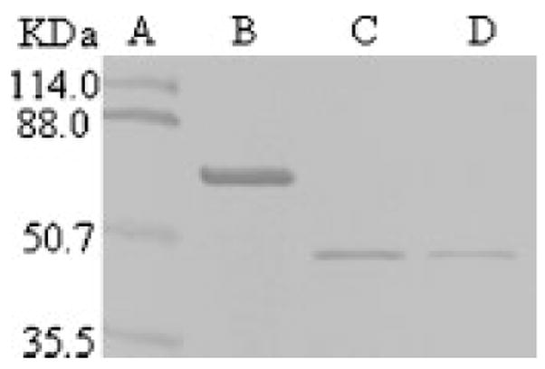
Expression, purification and thrombin cleavage of GST fused bovine osteopontin. Lane A: Prestained protein marker. Lane B: GST fused OPN, no thrombin. Lane C: The released recombinant OPN at 4 U thrombin per mg of GST fused protein. Lane D: The released recombinant OPN at 2 U thrombin per mg of GST fused protein. A portion of the recombinant protein was mixed with protein loading dye, fractionated onto 10% SDS-PAGE, and stained with Stains-all.
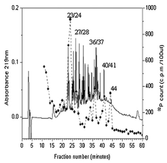
The phosphorylated tryptic peptides of recombinant bovine osteopontin. The peptides were separated by RP-HPLC on a Vydac C18 column (25 cm × 0.46 cm) using a linear gradient from H2O and 0.1% (v/v) trifluoroacetic acid to 60% CH3CN and 0.055% (v/v) trifluoroacetic acid over 80 min at a flow rate of 1 ml/min. Absorbance at 219 nm was recorded continuously. Fractions of 1 ml were collected, and aliquots were counted for 32P radioactivity. The phosphorylated tryptic peptides are numbered.
References
-
- Ashkar S, Weber GF, Panoutsakopoulou V, Sanchirico ME, Jansson M, Zawaideh S, Rittling SR, Denhardt DT, Glimcher MJ, Cantor H. Eta-1 (osteopontin): An early component of type-1 (cell-mediated) immunity. Science. 2000;287:860–864. - PubMed
-
- Carr C, McCount D, Cohen JB. The 43-kilodalton protein of Torpedo nicotinic postsynaptic membranes: Purification and determination of primary structure. Biochemistry. 1987;26:7090–7102. - PubMed
-
- Chen Y, Bal BS, Gorski JP. Calcium and collagen binding properties of osteopontin, bone sialoprotein, and bone acidic glycoprotein-75 from bone. J Biol Chem. 1992;267:24871–24878. - PubMed
-
- Chen J, Singh K, Mukherjee BB, Sodek J. Developmental expression of osteopontin (OPN) mRNA in rat tissues: Evidence for a role for OPN in bone formation and resorption. Matrix. 1993;13:113–123. - PubMed
-
- Christensen B, Nielsen MS, Haselmann KF, Petersen TE, Sorensen ES. Posttranslationally modified residues of native human osteopontin are located in clusters. Identification of thirty-six phosphorylation and five O-glycosylation sites and their biological implications. Biochem J. 2005;390:285–292. - PMC - PubMed
Publication types
MeSH terms
Substances
Grants and funding
LinkOut - more resources
Full Text Sources
Research Materials
