

Relationship between pulmonary surfactant protein and lipid peroxidation in lung injury due to paraquat intoxication in rats
- PMID: 17616020
- PMCID: PMC2687609
- DOI: 10.3904/kjim.2007.22.2.67
Relationship between pulmonary surfactant protein and lipid peroxidation in lung injury due to paraquat intoxication in rats
Abstract
Background: Pulmonary damage resulting from lipid peroxidation is a principal effect of paraquat intoxication. The host-defense functions of surfactant are known to be mediated by the surfactant proteins A and D (SP-A and SP-D, respectively). The primary objective of this study was to evaluate the variations over time in levels of surfactant protein and lipid peroxidation (LPO) in lung tissue following free-radical-induced injury.
Methods: 42 adult, male, Sprague-Dawley rats were administered intraperitoneal injections of paraquat (35 mg/kg body weight). SP-A and SP-D levels were determined via Western blot. LPO in the left lung homogenate was measured via analyses of the levels of thiobarbituric acid-reactive substances.
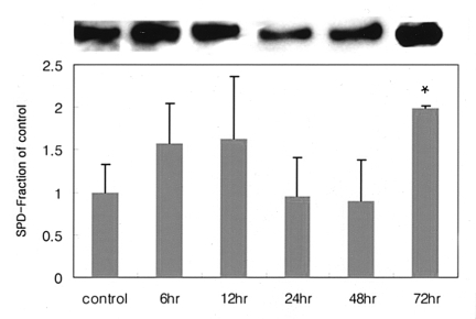
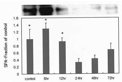
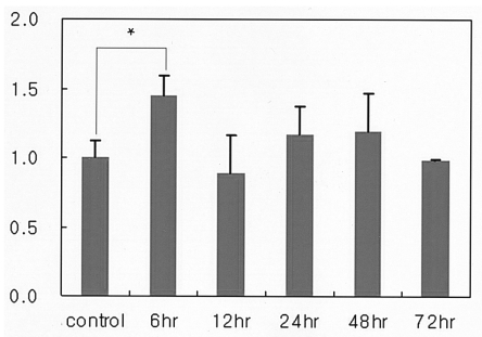
Results: LPO levels peaked at 6 hours, with no associated histological changes. SP-D levels increased until hour 12 and declined until hour 48; SP-D levels subsequently began to increase again, peaking at hour 72. SP-A levels peaked at hour 6, declining thereafter.
Conclusions: We suggest that in the early phase of paraquat injury, SP-D levels reflect alveolar damage and that de novo synthesis of SP-D takes 72 hours. Levels of SP-A, on the other hand, reflect abnormalities in the surfactant system in the late stage of paraquat intoxication. Surfactant proteins may play a role in protecting the lungs from reactive oxygen injury. A time-dependent variation has been observed in the levels of surfactant proteins A and D following paraquat injury, and it has been suggested that these proteins play a role in the protection of lung tissue against ROS-induced injuries.
Figures
Similar articles
-
[Expression of the pulmonary surfactant in rat with paraquat-induced acute lung injury].Zhonghua Yi Xue Za Zhi. 2015 Dec 8;95(46):3762-5. Zhonghua Yi Xue Za Zhi. 2015. PMID: 26850018 Chinese.
-
Methylprednisolone does not enhance the surfactant effects on oxygenation and histology in paraquat-induced rat lung injury.Intensive Care Med. 2002 Aug;28(8):1138-44. doi: 10.1007/s00134-002-1350-2. Epub 2002 Jun 13. Intensive Care Med. 2002. PMID: 12185438
-
Plasma surfactant D in patients following acute paraquat intoxication.Clin Toxicol (Phila). 2007 Jun-Aug;45(5):463-7. doi: 10.1080/15563650701338138. Clin Toxicol (Phila). 2007. PMID: 17503247
-
Pulmonary surfactant: an immunological perspective.Cell Physiol Biochem. 2010;25(1):13-26. doi: 10.1159/000272047. Epub 2009 Dec 22. Cell Physiol Biochem. 2010. PMID: 20054141 Free PMC article. Review.
-
Signaling pathways involved in paraquat-induced pulmonary toxicity: Molecular mechanisms and potential therapeutic drugs.Int Immunopharmacol. 2022 Dec;113(Pt A):109301. doi: 10.1016/j.intimp.2022.109301. Epub 2022 Oct 21. Int Immunopharmacol. 2022. PMID: 36279672 Review.
Cited by
-
Repeated Administration of Clinically Relevant Doses of the Prescription Opioids Tramadol and Tapentadol Causes Lung, Cardiac, and Brain Toxicity in Wistar Rats.Pharmaceuticals (Basel). 2021 Jan 27;14(2):97. doi: 10.3390/ph14020097. Pharmaceuticals (Basel). 2021. PMID: 33513867 Free PMC article.
-
Sex and SP-A2 Dependent NAD(H) Redox Alterations in Mouse Alveolar Macrophages in Response to Ozone Exposure: Potential Implications for COVID-19.Antioxidants (Basel). 2020 Sep 25;9(10):915. doi: 10.3390/antiox9100915. Antioxidants (Basel). 2020. PMID: 32992843 Free PMC article.
-
Sex differences in the acute in vivo effects of different human SP-A variants on the mouse alveolar macrophage proteome.J Proteomics. 2014 Aug 28;108:427-44. doi: 10.1016/j.jprot.2014.06.007. Epub 2014 Jun 18. J Proteomics. 2014. PMID: 24954098 Free PMC article.
-
Inhibitory effects of rosiglitazone on paraquat-induced acute lung injury in rats.Acta Pharmacol Sin. 2013 Oct;34(10):1317-24. doi: 10.1038/aps.2013.65. Epub 2013 Aug 12. Acta Pharmacol Sin. 2013. PMID: 23933652 Free PMC article.
-
The impact of surfactant protein-A on ozone-induced changes in the mouse bronchoalveolar lavage proteome.Proteome Sci. 2009 Mar 26;7:12. doi: 10.1186/1477-5956-7-12. Proteome Sci. 2009. PMID: 19323824 Free PMC article.
References
-
- Rose MS, Smith LL, Wyatt I. Evidence for energy-dependent accumulation of paraquat into rat lung. Nature. 1974;252:314–315. - PubMed
-
- Forman HJ, Aldrich TK, Posner MA, Fisher AB. Differential paraquat uptake and redox kinetics of rat granular pneumocytes and alveolar macrophages. J Pharmacol Exp Ther. 1982;221:428–433. - PubMed
-
- Winchester JF. Paraquat and the bipyridyl herbicides. In: Hadadad LM, Winchester JF, editors. Clinical management of poisoning and drug overdose. 2nd ed. Philadelphia: WB Saunders; 1990. pp. 1088–1103.
-
- Clark HW, Reid KB, Sim RB. Collectins and innate immunity in the lung. Microbes Infect. 2000;2:273–278. - PubMed
Publication types
MeSH terms
Substances
LinkOut - more resources
Full Text Sources