

Sclerosing encapsulating peritonitis (abdominal cocoon) after abdominal hysterectomy
- PMID: 17616031
- PMCID: PMC2687622
- DOI: 10.3904/kjim.2007.22.2.125
Sclerosing encapsulating peritonitis (abdominal cocoon) after abdominal hysterectomy
Abstract
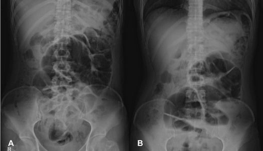
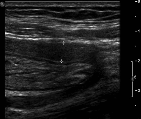
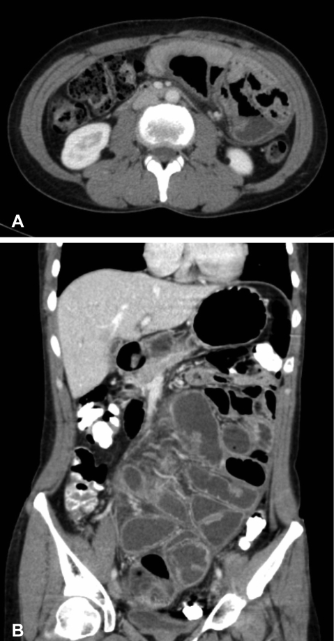
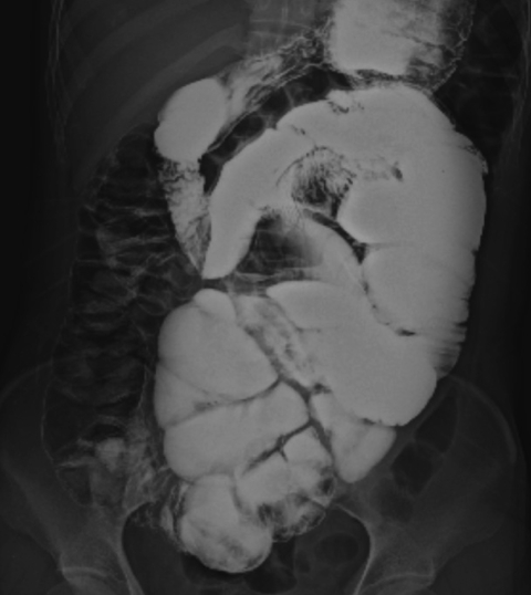
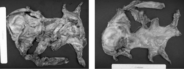
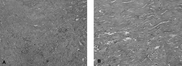
Sclerosing encapsulating peritonitis (SEP) is a poorly understood and rarely documented cause of small bowel obstruction. Although recurrent peritonitis has been reported as the main contributory factor leading to secondary SEP, the pathogenesis of primary (idiopathic) SEP is still uncertain. A 40-year-old woman with a history of total abdominal hysterectomy due to gestational trophoblastic disease presented with progressive lower abdominal pain and abdominal distension. Ultrasonography and contrast-enhanced abdomen-pelvis computed tomography of the abdomen revealed encapsulation of the entire small bowel with a sclerotic capsule. At laparotomy, a fibrous thick capsule encasing small bowel loops was revealed. Extensive adhesiolysis and removal of the capsule from the bowel loops were performed. The patient recovered uneventfully; she was discharged without complications. SEP is a rare cause of small bowel obstruction. We treated a case of abdominal cocoon with intestinal partial obstruction in a woman with a history of abdominal hysterectomy due to gestational trophoblastic disease. Surgical treatment was effective and the patient recovered without complication.
Figures
References
-
- Lee HY, Kim BS, Choi HY, Park HC, Kang SW, Choi KH, Ha SK, Han DS. Sclerosing encapsulating peritonitis as a complication of long term continuous ambulatory peritoneal dialysis in Korea. Nephrology. 2003;8(Suppl):S33–S39. - PubMed
-
- Kawaguchi Y, Kawaguchi H, Mujais S, Topley N, Oreopoulos DG. Encapsulating peritoneal sclerosis: definition, etiology, diagnosis, and treatment. Perit Dial Int. 2000;20(Suppl 4):S43–S55. - PubMed
-
- Foo KT, Ng KC, Rauff A, Foong WC, Sinniah R. Unusual small intestinal obstruction in adolescent girls: the abdominal cocoon. Br J Surg. 1978;65:427–430. - PubMed
-
- Marinho A, Adelusi B. The abdominal cocoon: a case report. Br J Obstet Gynaecol. 1980;87:249–250. - PubMed
-
- Pollock CA. Bloody ascites in a patient after transfer from peritoneal dialysis to hemodialysis. Semin Dial. 2003;16:406–410. - PubMed
Publication types
MeSH terms
LinkOut - more resources
Full Text Sources
Medical