

Myocardial fibrosis in a horse with polymorphic ventricular tachycardia observed during general anesthesia
- PMID: 17616061
- PMCID: PMC1876193
Myocardial fibrosis in a horse with polymorphic ventricular tachycardia observed during general anesthesia
Abstract
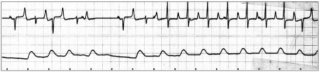
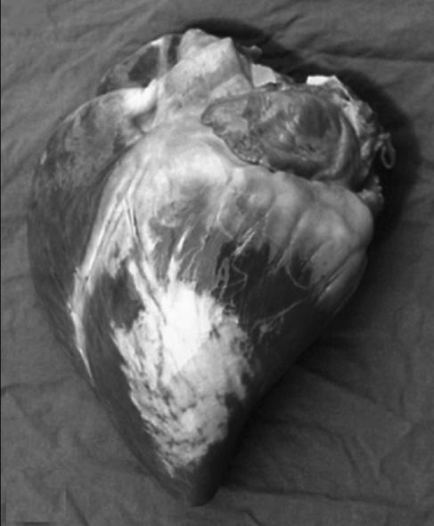
Ventricular dysrhythmias are more commonly associated with myocardial disease than are supraventricular dysrhythmias. Management of arrhythmias under general anesthesia is difficult because of the dysrhythmogenic effects of the anesthetic drugs. This report describes a severe ventricular dysrhythmia observed in a pony under general anesthesia, with a severe and old myocardial fibrosis found on postmortem examination.
Fibrose du myocarde chez un cheval présentant de la tachycardie ventriculaire polymorphe observée au cours d’une anesthésie générale. Les dysrythmies ventriculaires sont plus souvent associées aux maladies du myocarde que ne le sont les dysrythmies supraventriculaires. Les mesures à prendre lors d’arythmie survenant lors d’anesthésie générales sont compliquées par les effets dysrythmogènes des agents anesthésiques. Ce rapport décrit une dysrythmie ventriculaire grave observée chez un poney au cours d’une anesthésie générale et dont on a constaté à la nécropsie une grave et vielle fibrose du myocarde.
(Traduit par Docteur André Blouin)
Figures
Similar articles
-
ECG of the month.J Am Vet Med Assoc. 1993 Oct 1;203(7):972-3. J Am Vet Med Assoc. 1993. PMID: 8226263 No abstract available.
-
Anesthesia case of the month. Multiform ventricular premature contractions and nonsustained ventricular tachycardia during anesthesia.J Am Vet Med Assoc. 2010 Apr 15;236(8):840-3. doi: 10.2460/javma.236.8.840. J Am Vet Med Assoc. 2010. PMID: 20392178 No abstract available.
-
Ventricular tachycardia and myocardial dysfunction in a horse.J Am Vet Med Assoc. 1994 Dec 1;205(11):1569-73. J Am Vet Med Assoc. 1994. PMID: 7730126
-
Anesthesia and Myopathies of Horses.Vet Clin North Am Equine Pract. 2025 Apr;41(1):165-180. doi: 10.1016/j.cveq.2024.11.008. Epub 2024 Dec 30. Vet Clin North Am Equine Pract. 2025. PMID: 39741097 Review.
-
[Arrhythmogenic right ventricular disease].Z Kardiol. 1994;83 Suppl 6:175-80. Z Kardiol. 1994. PMID: 7863692 Review. German.
Cited by
-
The Health Status of Horses Used for at Least Six Complete Cycles of Loxoscelic Antivenom Production.Toxins (Basel). 2023 Sep 26;15(10):589. doi: 10.3390/toxins15100589. Toxins (Basel). 2023. PMID: 37888620 Free PMC article.
References
-
- McGuirk SM, Muir WW. Diagnosis and treatment of cardiac arrhythmias. Vet Clin North Am Equine Pract. 1985;1:353–370. - PubMed
-
- Muir WW. Anaesthetic complications and cardiopulmonary resuscitation in the horse. In: Reinhardt RW, Steube M, eds. Equine Anaesthesia: Monitoring and Emergency Therapy. St Louis: Mosby-Year Book, 1991: 461–484.
-
- Reimer JM, Reef VB, Sweeney RW. Ventricular arrhythmias in horses: 21 cases (1984–1989) J Am Vet Med Assoc. 1992;201:1237–1243. - PubMed
-
- Traub-Dargatz JL, Schlipf JW, Boon J, et al. Ventricular tachycardia and myocardial dysfunction in a horse. J Am Vet Med Assoc. 1994;205:1569–1573. - PubMed
-
- Schwarzwald CC, Hardy J, Buccellato M. High cardiac troponin I serum concentration in a horse with multiform ventricular tachycardia and myocardial necrosis. J Vet Intern Med. 2003;17:364–368. - PubMed
Publication types
MeSH terms
LinkOut - more resources
Full Text Sources