. 2007 Sep;189(17):6397-406.
doi: 10.1128/JB.00024-07. Epub 2007 Jul 6.

Structural characterization of the ribosome maturation protein, RimM

Affiliations

Structural characterization of the ribosome maturation protein, RimM

Sakura Suzuki et al. J Bacteriol. 2007 Sep.

Abstract

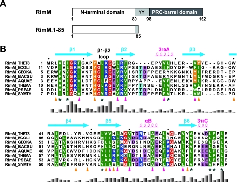
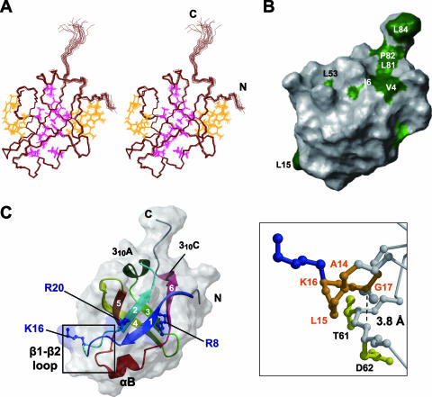
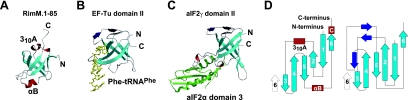
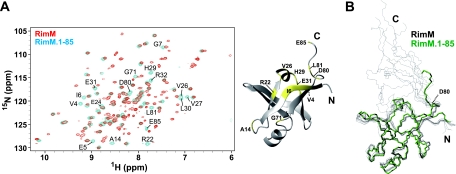
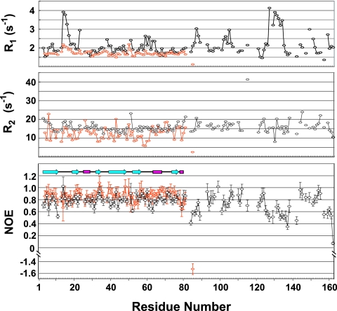
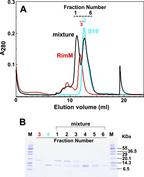
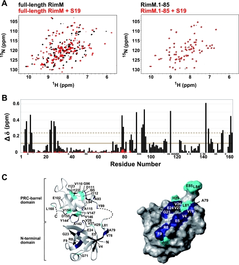
The RimM protein has been implicated in the maturation of the 30S ribosomal subunit. It binds to ribosomal protein S19, located in the head domain of the 30S subunit. Multiple sequence alignments predicted that RimM possesses two domains in its N- and C-terminal regions. In the present study, we have produced Thermus thermophilus RimM in both the full-length form (162 residues) and its N-terminal fragment, spanning residues 1 to 85, as soluble proteins in Escherichia coli and have performed structural analyses by nuclear magnetic resonance spectroscopy. Residues 1 to 80 of the RimM protein fold into a single structural domain adopting a six-stranded beta-barrel fold. On the other hand, the C-terminal region of RimM (residues 81 to 162) is partly folded in solution. Analyses of 1H-15N heteronuclear single quantum correlation spectra revealed that a wide range of residues in the C-terminal region, as well as the residues in the vicinity of a hydrophobic patch in the N-terminal domain, were dramatically affected upon complex formation with ribosomal protein S19.


Figures

FIG. 1.
FIG. 1.
(A) Predicted domain structure of the bacterial RimM proteins (top) and the RimM fragment used for our structural and biochemical studies (bottom). Residue numbers are for the RimM protein from T. thermophilus. The two conserved tyrosine residues are indicated by the one-letter amino acid code. (B) Amino acid sequence alignment of residues 1 to 85 in T. thermophilus RimM from different bacterial species. The positions of secondary structure elements, as observed in RimM.1-85 from T. thermophilus, are shown above the sequence. The side chains of the conserved hydrophobic residues involved in the core and on the side surfaces of the β-barrel are marked below with magenta or orange triangles, respectively. Conserved but solvent-exposed hydrophobic residues are marked with asterisks. Conserved glycine, hydrophobic, and aromatic residues are shown in red, green, or orange, respectively. Positively and negatively charged conserved residues are highlighted in blue or pink, respectively. The multiple sequence alignment was performed using ClustalX (7) and was adjusted manually to align the structurally significant residues. The histogram below the sequence indicates degrees of similarity. Species abbreviations: THET8, Thermus thermophilus HB8; SYMTH, Symbiobacterium thermophilum; AQUAE, Aquifex aeolicus; THEMA, Thermotoga maritima; GEOKA, Geobacillus kaustophilus; BACSU, Bacillus subtilis; ECOLI, Escherichia coli; PSEAE, Pseudomonas aeruginosa.
FIG. 2.
FIG. 2.
1H-15N HSQC spectrum of RimM.1-85, recorded at 25°C on a 600-MHz spectrometer. Signals are labeled with the residue number and the one-letter amino acid code.
FIG. 3.
FIG. 3.
Tertiary structure of RimM.1-85. (A) Stereo view illustrating a trace of the backbone atoms for the ensemble of the 20 structures with the lowest CYANA target function of RimM.1-85. Interior and exterior side chains that stabilize the RimM.1-85 structure are shown in magenta and orange, respectively. The colors of these side chains correspond to those of the triangles in Fig. 1B. (B) Mapping of hydrophobic residues on the molecular surface of RimM.1-85, generated by the MOLMOL (23) program. This structure is rotated by 60° around the y axis, as in panel A. Conserved hydrophobic residues among bacterial RimM proteins are colored green, and conserved but exposed hydrophobic residues are dark green. A large number of conserved hydrophobic residues are invisible, suggesting that they are deeply buried in the core. (C) Ribbon representation of the RimM.1-85 structure (left) in the same orientation as in panel A. The strands in the β-sheet are indicated by arrows, and the secondary structure elements are labeled. The side chains of the three conserved basic residues (blue) are linearly arranged on the surface of RimM.1-85. Right, enlarged view of the framed area of the left panel. The highly conserved segment in the β1-β2 loop, which has been regarded as the GXXG motif, is represented as an orange ball-and-stick model. Some of the residues relevant to the discussion are labeled. The broken line represents the distance between the α-carbon atom of G17 and the nitrogen atom of D62.
FIG. 4.
FIG. 4.
Comparison of interfaces in RIEF fold proteins. The β-sheets in the RIEF folds are shown in cyan and are depicted in the same orientation as the RimM.1-85 structure shown in Fig. 3C. The target nucleotides and proteins are partially displayed and are colored yellow and green, respectively. The characteristic structural elements in translation proteins are colored navy. (A) RimM.1-85. (B) Domain II of T. aquaticus EF-Tu with Phe-tRNAPhe (PDB identifier 1TTT) (31). (C) Domain II of archaeal aIF2γ with domain 3 of aIF2α (PDB identifier 2AHO) (37). (D) Topology diagrams of RimM.1-85 (left) and translation proteins (right). The six common strands are cyan, and the dissimilar secondary structures, namely helices and strands, are red and navy, respectively.
FIG. 5.
FIG. 5.
Superposition of full-length RimM and RimM.1-85. (A) Left, 1H-15N HSQC spectra of full-length RimM (red) and RimM.1-85 (blue), recorded at 25°C on a 600-MHz spectrometer. Most of the resonances originating from the N-terminal domain were overlapped with each other. Only the shifted resonances for residues 1 to 85 of full-length RimM, compared to those for RimM.1-85 itself, are labeled with the residue number and the one-letter amino acid code: V4, E5, I6, G7, A14, R22, E24, V26, V27, H29, L30, E31, R32, G71, D80, L81, and E85. Right, mapping of these residues (yellow) on the RimM.1-85 structure. (B) Superposition of the backbones of the 10 best structures of full-length RimM (black) and the best structure of RimM.1-85 (green). The 10 structures of full-length RimM were calculated with CYANA 1.0.8. The structure of the C-terminal region (residues 96 to 162) is not displayed, because it does not adopt a rigid tertiary structure.
FIG. 6.
FIG. 6.
NMR dynamic studies for full-length RimM and RimM.1-85. 15N R1 (top) and 15N R2 (middle) relaxation rates and steady-state 1H-15N NOE data (bottom) are shown for full-length RimM (black) and RimM.1-85 (orange), respectively. β-Strands, helices, and loops are indicated schematically.
FIG. 7.
FIG. 7.
Complex formation between full-length RimM and S19. (A) Size exclusion chromatography, performed using a Superdex 75 10/30 column. The mixture of full-length RimM and S19 in the molar ratio of 1:2 is shown by the black line. Each chromatography step with full-length RimM (20 nmol; red) and S19 (40 nmol; cyan) was performed under the same conditions. Fraction numbers are indicated in the panel with the corresponding colors. (B) SDS-polyacrylamide gel electrophoresis of fractions collected from size exclusion chromatography in panel A. Fraction numbers are indicated at the top: fractions 3 (red number), 4 (cyan), and 1 to 6 (black) correspond to full-length RimM, S19, and their mixture, respectively. Molecular weight standards are displayed as “M.”
FIG. 8.
FIG. 8.
NMR studies of full-length RimM and RimM.1-85 with S19. (A) 1H-15N HSQC spectra of free full-length RimM (black) and the complex with S19 (red) obtained at 45°C (left) and spectra of free RimM.1-85 (black) and its 1:1 molar mixture with S19 (red) monitored at 45°C (right). (B) Chemical shift changes of full-length RimM (black bars) and RimM.1-85 (red bars) observed by the addition of S19. The chemical shift change, Δδ, was determined as follows: Δδ = [(ΔδHN)2 + (ΔδN/6.5)2]1/2 (29), where ΔδHN and ΔδN are the chemical shift differences for HN and 15N, respectively. The mean value is shown by a continuous line; the mean value plus 1 standard deviation is shown by a dashed line. Asterisks indicate residues with 1H-15N resonances that were not assigned in the S19-free or S19-bound form of full-length RimM. (C) Mapping of perturbed residues on the homology-modeled structure of full-length RimM (left) and the surface of RimM.1-85, which is presented in the same orientation as in Fig. 3B (right). The 3D model of full-length RimM from T. thermophilus was obtained using the homology modeling approach with the SWISS-MODEL Protein Modeling Server (http://swissmodel.expasy.org/) (36) on the basis of the crystal structure of P. aeruginosa RimM. Disordered regions in the crystal structure are depicted by a dashed line. Residues with chemical shift changes above average are cyan, and residues with chemical shift changes above the mean value plus 1 standard deviation are navy.

Similar articles

Cited by

References

    1. Alix, J. H. 1993. Extrinsic factors in ribosome assembly, p. 173-184. In K. H. Nierhaus, F. Franceschi, and A. R. Subramanian (ed.), The translational apparatus: structure, function, regulation, evolution. Plenum, New York, NY.
    1. Allen, G. S., A. Zavialov, R. Gursky, M. Ehrenberg, and J. Frank. 2005. The cryo-EM structure of a translation initiation complex from Escherichia coli. Cell 121:703-712. - PubMed
    1. Anantharaman, V., and L. Aravind. 2002. The PRC-barrel: a widespread, conserved domain shared by photosynthetic reaction center subunits and proteins of RNA metabolism. Genome Biol. 3:RESEARCH0061.1-RESEARCH0061.9. - PMC - PubMed
    1. Bylund, G. O., B. C. Persson, L. A. Lundberg, and P. M. Wikström. 1997. A novel ribosome-associated protein is important for efficient translation in Escherichia coli. J. Bacteriol. 179:4567-4574. - PMC - PubMed
    1. Bylund, G. O., L. C. Wipemo, L. A. Lundberg, and P. M. Wikström. 1998. RimM and RbfA are essential for efficient processing of 16S rRNA in Escherichia coli. J. Bacteriol. 180:73-82. - PMC - PubMed

Publication types

Associated data

LinkOut - more resources