. 2007 Jul 1;67(13):6192-203.
doi: 10.1158/0008-5472.CAN-06-4424.

Retinoblastoma tumor suppressor status is a critical determinant of therapeutic response in prostate cancer cells

Affiliations

Retinoblastoma tumor suppressor status is a critical determinant of therapeutic response in prostate cancer cells

Ankur Sharma et al. Cancer Res. .

Abstract

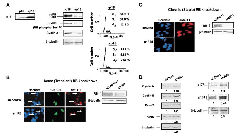
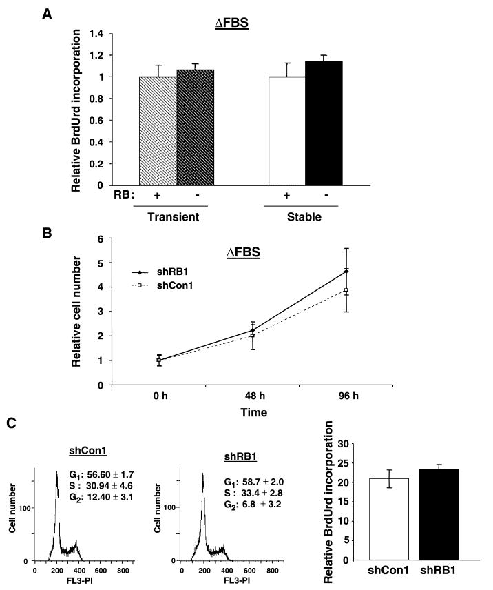
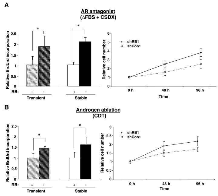
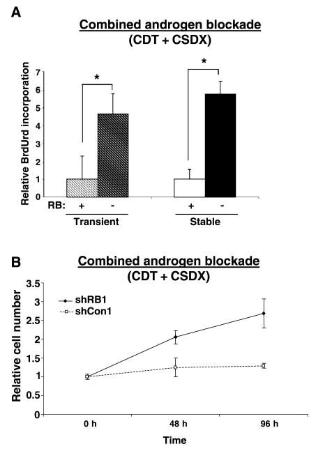
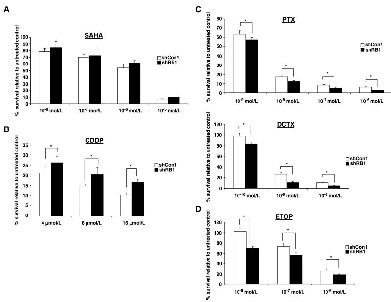
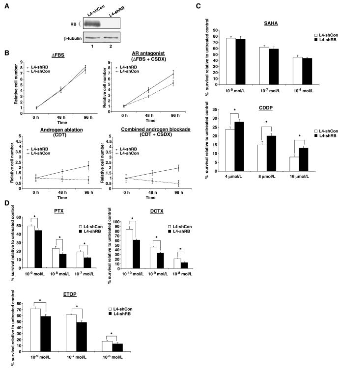
The retinoblastoma tumor suppressor protein (RB), a critical mediator of cell cycle progression, is functionally inactivated in the majority of human cancers, including prostatic adenocarcinoma. The importance of RB tumor suppressor function in this disease is evident because 25% to 50% of prostatic adenocarcinomas harbor aberrations in RB pathway. However, no previous studies challenged the consequence of RB inactivation on tumor cell proliferation or therapeutic response. Here, we show that RB depletion facilitates deregulation of specific E2F target genes, but does not confer a significant proliferative advantage in the presence of androgen. However, RB-deficient cells failed to elicit a cytostatic response (compared with RB proficient isogenic controls) when challenged with androgen ablation, AR antagonist, or combined androgen blockade. These data indicate that RB deficiency can facilitate bypass of first-line hormonal therapies used to treat prostate cancer. Given the established effect of RB on DNA damage checkpoints, these studies were then extended to determine the impact of RB depletion on the response to cytotoxic agents used to treat advanced disease. In this context, RB-deficient prostate cancer cells showed enhanced susceptibility to cell death induced by only a selected subset of cytotoxic agents (antimicrotubule agents and a topoisomerase inhibitor). Combined, these data indicate that RB depletion dramatically alters the cellular response to therapeutic intervention in prostate cancer cells and suggest that RB status could potentially be developed as a marker for effectively directing therapy.


Figures

Figure 1
Figure 1
RB knockdown in prostate cancer cells causes deregulation of RB/E2F target genes and compensation by RB family members. A, LNCaP cells were infected with p16INK4a adenovirus. Following culture for 36 to 48 h, cells were harvested, lysed, and immunoblotted as indicated. β-Tubulin is included as the loading control (left and middle). p16INK4a-infected cells were analyzed in parallel for cell cycle position (right). B, cells were transfected with pMSCV-puroRb3C or pMSCV-puro and H2B-GFP as a marker for transfection. Transfected cells were visualized for RB staining by indirect immunofluorescence (40× magnification; left), and corresponding lysates were monitored for RB expression levels by immunoblot (right). C, stable clones of RB knockdown (shRB1) or control (shCon1) were generated and tested for RB expression levels using immunofluorescence (40× magnification; left) and immunoblot analysis (right). D, immunoblot analysis of shRB1 and shCon1 cells to monitor alteration of E2F targets (cyclin A, cyclin E, Mcm-7, PCNA). β-Tubulin is included as a loading control (left). Relative expression in shRB1 (normalized to β-tubulin and compared with shCon1) is indicated as determined by LICOR fluorescence. Lysates from shRB1 and shCon1 cells were subjected to immunoblot analyses to examine p107 and p130. β-Tubulin served as a loading control, and relative expression is indicated (right).
Figure 2
Figure 2
RB depletion confers no proliferative advantage in the presence of androgen. A, LNCaP cells transiently transfected with pMSCV-puroRb3C or pMSCV-puro (hashed columns) or stable clones described in Fig. 1B, shRB1 and shCon1 (solid columns) were cultured in ΔFBS and monitored for BrdUrd incorporation as described in Materials and Methods. For comparison, BrdUrd incorporation in each RB control was set to 1. B, shRB1 and shCon1 cells were seeded at equal density in the presence of androgen (ΔFBS), and after 24 h of culture (time, 0 h), cell number was monitored at the times indicated. Data represent at least three independent experiments wherein data for each time point were collected with triplicate biological replicates (P > 0.05, ANOVA). C, asynchronous populations of shRB1 and shCon1 cells were cultured for 48 h, pulsed with BrdUrd for 1 h, and then analyzed for cell cycle and BrdUrd incorporation. Data represent four independent samples (P > 0.05, ANOVA).
Figure 3
Figure 3
RB depletion reduces the efficacy of single agent–based hormonal intervention. A, as in Fig. 2, transient RB knockdown cells or stable clones were cultured in androgen containing media supplemented with 1 μmol/L Casodex or 0.1% DMSO (vehicle control). After 48 h of treatment, cells were then monitored for relative BrdUrd incorporation (left), or cell number was monitored at indicated time points (right). B, experiments were repeated as in (A), except that cells were cultured in steroid-depleted serum (CDT) for 48 h before BrdUrd labeling (left) or monitored for cell viability as indicated (right). Data represent at least three independent experiments wherein data for each time point were collected with triplicate biological replicates. *, P < 0.05, ANOVA.
Figure 4
Figure 4
RB depletion triggers bypass of combined androgen blockade. As in Fig. 3B, transient RB knockdown cells or stable clones were cultured in steroid-depleted serum (CDT) media supplemented with 1 μmol/L Casodex or 0.1% DMSO (vehicle control). After 48 h of treatment, cells were then monitored for relative BrdUrd incorporation (A) or cell number monitored at indicated time points (B). Data represent at least three independent experiments, wherein data for each time point were collected with triplicate biological replicates. *, P < 0.05, ANOVA.
Figure 5
Figure 5
RB depletion sensitizes prostate cancer cells to selected chemotherapeutics. Stable clones were cultured in androgen-containing media (ΔFBS), and following 24 h in culture, cells were supplemented with indicated concentrations of SAHA (A), cisplatin (CDDP; B), antimicrotubule agents paclitaxel (PTX) and docetaxel (DCTX; C), and etoposide (ETOP; D) or 0.1% DMSO (vehicle control) and challenged for 48 h. Cells were resupplemented with fresh media without drugs and allowed to propagate for another 48 h and then counted using trypan blue exclusion method. The number of cells remaining after drug treatment was set relative to condition without cytotoxic challenge (100% survival). Data represent at least three independent experiments, wherein data were collected with duplicate biological replicates. *, P < 0.05, ANOVA.
Figure 6
Figure 6
Impact of RB depletion is conserved in multiple prostate cancer cell systems. A, stable clones of RB knockdown (L4-shRB) or control (L4-shCon) were generated from parental LAPC-4 cells and tested for RB expression levels using immunoblot analysis. B, L4-shRB and L4-shCon cells were cultured in ΔFBS or CDT media supplemented with or without Casodex, and cell number was monitored at indicated time points using trypan blue exclusion. Data represent at least three independent experiments wherein data for each time point were collected with triplicate biological replicates. C and D, stable clones were cultured in ΔFBS, treated with SAHA, cisplatin, paclitaxel, docetaxel, and etoposide as described in Fig. 5, and cell number was measured using trypan blue exclusion. The number of cells remaining after drug treatment was set relative to condition without cytotoxic challenge (100% survival). Data represent at least three independent experiments wherein data were collected with duplicate biological replicates. *, P < 0.05, ANOVA.

Similar articles

Cited by

References

    1. Jemal A, Murray T, Ward E, et al. Cancer statistics, 2005. CA Cancer J Clin. 2005;55:10–30. - PubMed
    1. Kolvenbag GJ, Nash A. Bicalutamide dosages used in the treatment of prostate cancer. Prostate. 1999;39:47–53. - PubMed
    1. Sharifi N, Gulley JL, Dahut WL. Androgen deprivation therapy for prostate cancer. JAMA. 2005;294:238–44. - PubMed
    1. Feldman BJ, Feldman D. The development of androgen-independent prostate cancer. Nat Rev Cancer. 2001;1:34–45. - PubMed
    1. Miyamoto H, Messing EM, Chang C. Androgen deprivation therapy for prostate cancer: current status and future prospects. Prostate. 2004;61:332–53. - PubMed

Publication types