. 2007 Jul;19(7):2246-63.
doi: 10.1105/tpc.106.042697. Epub 2007 Jul 6.

The TORMOZ gene encodes a nucleolar protein required for regulated division planes and embryo development in Arabidopsis

Affiliations

The TORMOZ gene encodes a nucleolar protein required for regulated division planes and embryo development in Arabidopsis

Megan E Griffith et al. Plant Cell. 2007 Jul.

Abstract

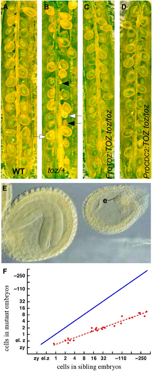
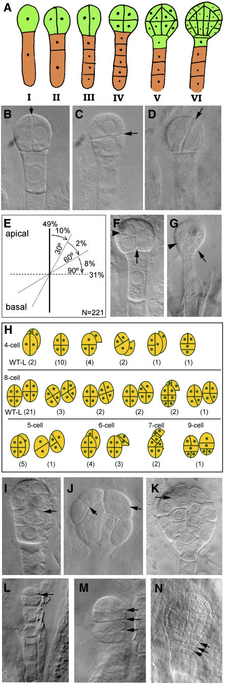
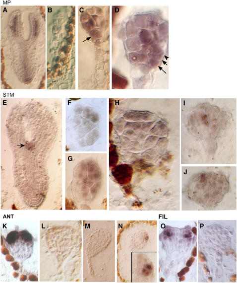
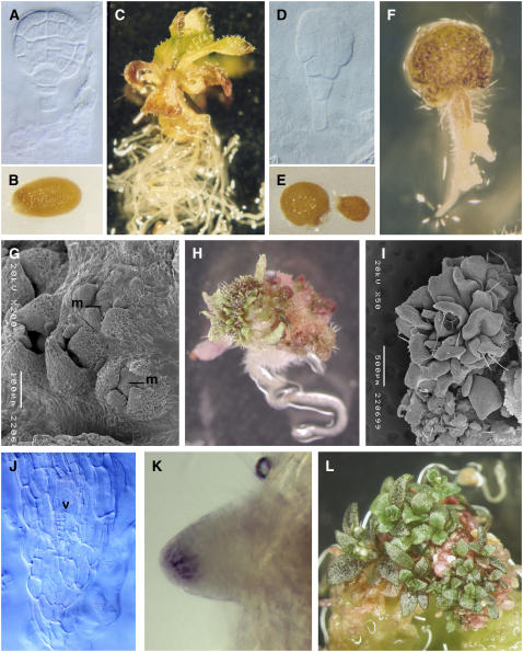
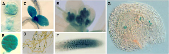
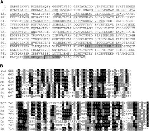
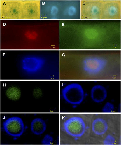
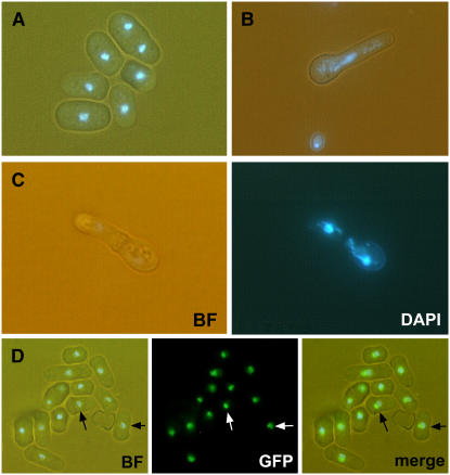
Embryogenesis in Arabidopsis thaliana is marked by a predictable sequence of oriented cell divisions, which precede cell fate determination. We show that mutation of the TORMOZ (TOZ) gene yields embryos with aberrant cell division planes and arrested embryos that appear not to have established normal patterning. The defects in toz mutants differ from previously described mutations that affect embryonic cell division patterns. Longitudinal division planes of the proembryo are frequently replaced by transverse divisions and less frequently by oblique divisions, while divisions of the suspensor cells, which divide only transversely, appear generally unaffected. Expression patterns of selected embryo patterning genes are altered in the mutant embryos, implying that the positional cues required for their proper expression are perturbed by the misoriented divisions. The TOZ gene encodes a nucleolar protein containing WD repeats. Putative TOZ orthologs exist in other eukaryotes including Saccharomyces cerevisiae, where the protein is predicted to function in 18S rRNA biogenesis. We find that disruption of the Sp TOZ gene results in cell division defects in Schizosaccharomyces pombe. Previous studies in yeast and animal cells have identified nucleolar proteins that regulate the exit from M phase and cytokinesis, including factors involved in pre-rRNA processing. Our study suggests that in plant cells, nucleolar functions might interact with the processes of regulated cell divisions and influence the selection of longitudinal division planes during embryogenesis.


Figures

Figure 1.
Figure 1.
Developing Seed Phenotypes. (A) and (B) Superficial phenotypes of seeds from wild-type (A) and toz/+ (B) siliques. The closed arrows highlight aborted seeds, and the open arrows highlight living seeds with reduced rates of development. (C) and (D) Siliques from toz/toz plants complemented with a genomic fragment (ProTOZ:TOZ) still produce a few aborted seed (C), whereas the toz/toz siliques complemented by the TOZ cDNA driven by the CDC2a promoter (ProCDC2:TOZ) show very few aborted embryos (D). (E) Late development of embryos in a single silique, comparing the toz mutants (e) to the sibling embryo at an advanced stage. (F) The comparison of growth rates, where each point on the graph represents the average number of cells in mutant versus sibling embryos within a single silique. The solid blue line shows the expected positions of the points if there were a 1:1 correspondence between cell divisions in the wild type versus toz mutant embryos.
Figure 2.
Figure 2.
Early Embryo Development. (A) Wild-type embryos follow a predictable pattern of cell division. The stages represent sections of one-cell (I), two-cell (II), four-cell (III), eight-cell (IV), 16-cell (V), and 32-cell (VI) proembryos (green). (B) to (D) Whole-mount cleared toz embryos showing the first division of the apical cell (arrows) with cell divisions that are longitudinal as in the wild type (B), transverse (C), or oblique (D). (E) The division planes observed in two-cell proembryos with angles relative to the apical-basal axis are illustrated. (F) A toz proembryo with three cells. The first division was longitudinal (arrow) and the second transverse (arrowhead). (G) A four-cell proembryo. The first division was oblique (arrow); the left cell then divided transversely relative to the right cell wall (arrowhead). The right cell divided in the plane of the paper such that only one cell is visible. (H) Illustration of the variety of embryos and associated division patterns observed in toz proembryos when sibling embryos were at the heart stage. The pairs of nearly superimposed circles, or those offset to the right of each diagram, represent daughters of a division that occurred in the plane of the page. The numbers in parentheses give the numbers observed for each pattern, with the left-most illustration representing toz embryos exhibiting the wild-type-like pattern (indicated by WT-L). (I) to (K) Multicell toz embryos at later stages, with arrows highlighting unpredictable divisions. The embryo in (I) consists of six cells (the upper left cell overlays another cell that is outside the plane of view). toz cells can be large (J) and give rise to globular embryos with atypical cell patterning (K). (L) to (N) Phenotypes of embryos from transgenic lines harboring the TOZ antisense cDNA driven by the At CDC2 promoter. Transverse divisions that replace longitudinal divisions were observed in two-cell proembryos (L). Transverse divisions absent in nontransformed controls were frequently observed in older embryos ([M] and [N], arrows).
Figure 3.
Figure 3.
Gene Expression Patterns in TOZ Embryos. Analysis of gene expression of MP ([A] to [D]), STM ([E] to [J]), ANT ([K] to [N]), and FIL ([O] and [P]). (A) Sibling embryo of wild-type phenotype expresses MP in vascular tissue and root tip as previously described (Hamann et al., 2002). (B) to (D) In toz embryos, the MP signal may be absent (B) or expressed nonuniformly ([C] and [D]). The cells in the presumptive hypophysis may continue to express MP (C), and cells in the outermost layer may also express MP ([D], arrows). (E) STM expression marks the shoot apical meristem in wild-type cells (arrow). (F) to (H) In mutant cells, STM is widely expressed across a number of apically positioned cells. (I) and (J) In an older embryo, STM is confined to a few cells in the subdermal or epidermal layers as shown by consecutive sections. (K) and (L) In the wild type, ANT is first detected at the late globular stage, just prior to the outgrowth of the lateral organ primordial (K). However, in toz embryos, which consist of many more cells, no ANT expression was observed (L). (M) and (N) It was not until a very late stage that a few cells showed ANT signal, as depicted by sections 1 and 4 of the same embryo (inset). (O) and (P) FIL expression in the wild type (O) is absent in the mutant embryo (P).
Figure 4.
Figure 4.
Phenotypes of ex Planta–Cultured Embryos. Wild-type ([A] to [C]) and toz ([D] to [L]) tissue were propagated by in vitro culture from early stages. (A) to (E) Wild-type embryos were cultured from preglobular, globular (A), or older stages. Most often wild-type embryos resulted in normal seed (B) and seedlings after 3 weeks (C). Mutant embryos (D) were cultured from several stages but with similar results. Sometimes the seed failed to germinate (right seed in [E]) or was abnormally shaped (left seed in [E]) compared with the wild type (B). (F) to (I) Approximately 2 weeks after germination, the apical region becomes a green mass of cells (F) that often gives rise to lateral organs ([H] and [L]). Developmental progression was observed by scanning electron microscopy of toz plants and revealed small centers of young lateral organ primordia developing around a central mound (m) of cells (G), which apparently induces prolific leaf production ([H] and [I]). (J) Whole-mount cleared sections of a primary root show that the vasculature (v) forms in the center of the root as in the wild type, whereas the root apex lacks regular no cell patterning. (K) Lugol staining detected the presence of amyloplasts in columella cells of some later toz roots. (L) Four to six weeks after germination, toz plantlets showed prolific vegetative tissue on a mass of cells, but little root tissue was observed.
Figure 5.
Figure 5.
TOZ Expression Patterns. In toz/+ heterozygous plants, GUS is expressed weakly at the four-cell proembryo stage (A) but strongly at the mid-globular stage (B). In the seedling, GUS staining is predominant in the shoot apex, young leaves (C), and root tips (D). In the inflorescence, the precipitate is strongest in the young stamens and carpels. Using a different staining solution that restricts the diffusion of intermediates, GUS can be localized to the nucleus in the roots (G) and also in each of the cells of the embryo sac in the mature ovule (H).
Figure 6.
Figure 6.
Molecular Analysis of the TOZ Gene. (A) The predicted translation from the cDNA sequence. The 12 WD40 repeat sequences have been underlined, the two putative nuclear localization signals are shaded, and the boxed amino acids represent those deleted in the mutant toz protein. (B) Comparative amino acid interspecies homology for the TOZ domain at the C-terminal region. Os, Oryza sativa; Hs, Homo sapiens; Mm, Mus musculus; Ce, Caenorhabditis elegans; Sc, Saccharomyces cerevisiae; Sp, Saccharomyces pombe. A full list of TOZ homologs with accession numbers is available in Supplemental Table 1 online.
Figure 7.
Figure 7.
Subcellular Localization of TOZ. (A) to (G) Root cells from toz/+ plants. Cells yielding blue precipitate (A) were counterstained with DAPI (B). The overlay shows that the GUS is predominantly restricted from the chromatin region of the nucleus (C). Immunofluorescence staining was used to confirm whether toz-GUS was restricted to the nucleolus. Signal from antibodies to the nucleolar-localized human fibrillarin gene is shown in red (D), in the toz-GUS protein (E), and by DAPI staining (F). The overlay (G) shows that the fibrillarin signal mostly colocalized with toz-GUS, yet both are excluded from the chromatin region of the nucleus. (H) to (K) Localization of TOZ-GFP fusion protein in roots. GFP fluorescence (H) localizes to the nonchromatin region of the nucleus where the nucleolus is located, as revealed by DAPI staining (I), DAPI and GFP (J), and DAPI, GFP, and bright-field images (K). The slight offset of the GFP signal relative to the DAPI signal is due to displacements in the focal planes of the two lasers at higher magnifications.
Figure 8.
Figure 8.
Analysis of Sp TOZ in S. pombe. (A) and (B) are superimposed bright-field and DAPI images. (C) shows bright-field and DAPI as separate panels. (D) shows superposed bright-field and DAPI images (left) as well as GFP (center) and merged images (right). (A) Wild-type pombe have distinct small nuclei as shown by DAPI staining. (B) and (C) By contrast, the deletion mutants (ΔSptoz) show germinated spores with dispersed nuclei. Some have undergone division, but the newly formed septum cuts the lagging DNA strands (C). (D) GFP was engineered to produce a translational fusion with Sp TOZ and transformed into wild-type pombe for homologous recombination at the Sp TOZ locus. Sp TOZ:GFP detection shows close proximity to the nonchromatin region of the S. pombe nucleus (arrows).

Similar articles

Cited by

References

    1. Andersen, J.S., Lyon, C.E., Fox, A.H., Leung, A.K., Lam, Y.W., Steen, H., Mann, M., and Lamond, A.I. (2002). Directed proteomic analysis of the human nucleolus. Curr. Biol. 12 1–11. - PubMed
    1. Apuya, N.R., Yadegari, R., Fischer, R.L., Harada, J.J., Zimmerman, L., and Goldberg, R.B. (2001). The Arabidopsis embryo mutant schlepperless has a defect in the chaperonin-60α gene. Plant Physiol. 126 717–730. - PMC - PubMed
    1. Barneche, F., Steinmetz, F., and Echeverria, M. (2000). Fibrillarin genes encode both a conserved nucleolar protein and a novel small nucleolar RNA involved in ribosomal RNA methylation in Arabidopsis thaliana. J. Biol. Chem. 275 27212–27220. - PubMed
    1. Basi, G., Schmid, E., and Maundrell, K. (1993). TATA box mutations in the Schizosaccharomyces pombe nmt1 promoter affect transcription efficiency but not the transcription start point or thiamine repressibility. Gene 123 131–136. - PubMed
    1. Baud, S., Bellec, Y., Miquel, M., Bellini, C., Caboche, M., Lepiniec, L., Faure, J.-D., and Rochat, C. (2004). gurke and pasticcino3 mutants affected in embryo development are impaired in acetyl-CoA carboxylase. EMBO Rep. 5 515–520. - PMC - PubMed

Publication types

MeSH terms