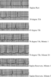
Dynamic changes in T-wave amplitude during tilt table testing: correlation with outcomes
- PMID: 17617070
- PMCID: PMC6932404
- DOI: 10.1111/j.1542-474X.2007.00168.x
Dynamic changes in T-wave amplitude during tilt table testing: correlation with outcomes
Abstract
Background: Changes in autonomic tone may play a role in syncope. Autonomic tone has been shown to affect cardiac repolarization in the ECG. Changes in the T wave can be seen during head-up tilt table (HUT) testing with unknown significance or relationship to outcomes.
Methods: Twelve-lead ECGs during HUT testing from 150 patients were reviewed from a prospectively collected registry database. ECGs during supine-rest, 30-45-70 degrees tilt, and 5-minute supine recovery were reviewed. Changes in the T wave, that is, decreased amplitude with or without becoming negative or flipping from negative to positive, were recorded for each stage. Outcomes of the HUT test include nondiagnostic, postural orthostatic hypotension (POH), postural orthostatic tachycardia syndrome (POTS), and vasovagal response (VVR). Age (Younger: <50 year old; Older: > or = 50 year old) and gender were analyzed.
Results: Of 150 patients (108 women; 80 Younger), 135 had T-wave changes during HUT; changes resolved in 114 patients during supine recovery. Changes mostly occurred in inferior and anterolateral leads. POH occurred in 114 patients, POTS in 67, and VVR in 30. T-wave changes in V1 inversely correlated with POH (P = 0.005). T-wave changes in inferior leads II, III, aVF and in anterolateral leads V3-V6 positively correlated with POTS (P < 0.05). Female gender and younger age correlated with POTS independent of the leads (P < 0.05). Concomitant T-wave changes in V5 and V6 correlated with VVR; changes in aVF also correlated with VVR (P < 0.05).
Conclusions: Dynamic T-wave changes during HUT testing in inferior and anterolateral leads are associated with POTS and VVR independent of age and gender. Changes in autonomic tone may play a role and need further study.
Figures


Similar articles
-
Dynamic changes of P-wave duration and P-wave axis during head-up tilt test in patients with vasovagal syncope.Pacing Clin Electrophysiol. 2006 Jul;29(7):742-6. doi: 10.1111/j.1540-8159.2006.00428.x. Pacing Clin Electrophysiol. 2006. PMID: 16884510
-
Diagnostic and prognostic value of T-wave amplitude difference between supine and orthostatic electrocardiogram in children and adolescents with postural orthostatic tachycardia syndrome.Ann Noninvasive Electrocardiol. 2020 Jul;25(4):e12747. doi: 10.1111/anec.12747. Epub 2020 Feb 29. Ann Noninvasive Electrocardiol. 2020. PMID: 32112609 Free PMC article.
-
High incidence of ST-T changes in women during tilt-table testing.J Electrocardiol. 2017 Nov-Dec;50(6):884-888. doi: 10.1016/j.jelectrocard.2017.06.004. Epub 2017 Jun 9. J Electrocardiol. 2017. PMID: 28645449
-
The effect of head-upright tilt table testing for vasovagal syncope on P-wave duration.J Electrocardiol. 2002 Oct;35(4):303-6. doi: 10.1054/jelc.2002.35847. J Electrocardiol. 2002. PMID: 12395356
-
[Clinical significance of changes in T wave and ST segment amplitudes on electrocardiogram from supine to standing position among children with unexplained chest tightness or pain in resting stage].Zhongguo Dang Dai Er Ke Za Zhi. 2013 Sep;15(9):771-4. Zhongguo Dang Dai Er Ke Za Zhi. 2013. PMID: 24034923 Chinese.
Cited by
-
Correlation analysis of deep learning methods in S-ICD screening.Ann Noninvasive Electrocardiol. 2023 Jul;28(4):e13056. doi: 10.1111/anec.13056. Epub 2023 Mar 15. Ann Noninvasive Electrocardiol. 2023. PMID: 36920649 Free PMC article.
-
The Repolarization Period during the Head-Up Tilt Test in Children with Vasovagal Syncope.Int J Environ Res Public Health. 2020 Mar 15;17(6):1908. doi: 10.3390/ijerph17061908. Int J Environ Res Public Health. 2020. PMID: 32183460 Free PMC article.
-
ST-segment changes during tilt table testing for postural tachycardia syndrome: correlation with exercise stress test results.Clin Auton Res. 2020 Feb;30(1):79-83. doi: 10.1007/s10286-019-00633-9. Epub 2019 Aug 21. Clin Auton Res. 2020. PMID: 31435848
-
Prolongation of Electrocardiographic T Wave Parameters Recorded during the Head-Up Tilt Table Test as Independent Markers of Syncope Severity in Children.Int J Environ Res Public Health. 2020 Sep 4;17(18):6441. doi: 10.3390/ijerph17186441. Int J Environ Res Public Health. 2020. PMID: 32899625 Free PMC article.
-
Peaked T--waves and sinus arrhythmia before prolonged sinus pauses and atrioventricular block in guillain-barre syndrome.Indian Pacing Electrophysiol J. 2007 Oct 22;7(4):249-52. Indian Pacing Electrophysiol J. 2007. PMID: 17957274 Free PMC article. No abstract available.
References
-
- Benditt DG, Ermis C, Padanilam B, et al Catecholamine response during haemodynamically stable upright posture in individuals with and without tilt‐table induced vasovagal syncope. Europace 2003;5:65–70. - PubMed
-
- Goldstein DS, Holmes C, Frank SM, et al Sympathoadrenal imbalance before neurocardiogenic syncope. Am J Cardiol 2003;91:53–58. - PubMed
-
- Lehmann KG, Shandling AH, Yusi AU, et al Altered ventricular repolarization in central sympathetic dysfunction associated with spinal cord injury. Am J Cardiol 1989;63:1498–1504. - PubMed
-
- Miyazaki T, Mitamura H, Miyoshi S, et al Autonomic and antiarrhythmic drug modulation of ST segment elevation in patients with Brugada syndrome. J Am Coll Cardiol 1996;27:1061–1070. - PubMed
MeSH terms
LinkOut - more resources
Full Text Sources

