

Characterization of a small plaque variant of West Nile virus isolated in New York in 2000
- PMID: 17617432
- PMCID: PMC2190729
- DOI: 10.1016/j.virol.2007.06.008
Characterization of a small plaque variant of West Nile virus isolated in New York in 2000
Abstract
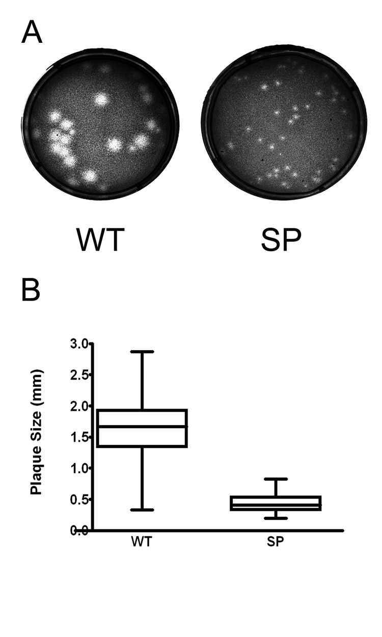
A small-plaque variant (SP) of West Nile virus (WNV) was isolated in Vero cell culture from kidney tissue of an American crow collected in New York in 2000. The in vitro growth of the SP and parental (WT) strains was characterized in mammalian (Vero), avian (DF-1 and PDE), and mosquito (C6/36) cells. The SP variant replicated less efficiently than did the WT in Vero cells. In avian cells, SP growth was severely restricted at high temperatures, suggesting that the variant is temperature sensitive. In mosquito cells, growth of SP and WT was similar, but in vivo in Culex pipiens (L.) there were substantial differences. Relative to WT, SP exhibited reduced replication following intrathoracic inoculation and lower infection, dissemination, and transmission rates following oral infection. Analysis of the full length sequence of the SP variant identified sequence differences which led to only two amino acid substitutions relative to WT, prM P54S and NS2A V61A.
Figures
Similar articles
-
West Nile Virus Activity in a Winter Roost of American Crows (Corvus brachyrhynchos): Is Bird-To-Bird Transmission Important in Persistence and Amplification?J Med Entomol. 2015 Jul;52(4):683-92. doi: 10.1093/jme/tjv040. Epub 2015 Apr 24. J Med Entomol. 2015. PMID: 26335475 Free PMC article.
-
Avian virulence and thermostable replication of the North American strain of West Nile virus.J Gen Virol. 2006 Dec;87(Pt 12):3611-3622. doi: 10.1099/vir.0.82299-0. J Gen Virol. 2006. PMID: 17098976
-
Genetic and phenotypic variation of West Nile virus in New York, 2000-2003.Am J Trop Med Hyg. 2004 Oct;71(4):493-500. Am J Trop Med Hyg. 2004. PMID: 15516648
-
The Spread of the Mosquito-Transmitted West Nile Virus in North America and Europe.Microb Biotechnol. 2025 Mar;18(3):e70120. doi: 10.1111/1751-7915.70120. Microb Biotechnol. 2025. PMID: 40035176 Free PMC article. Review.
-
Globalization, land use, and the invasion of West Nile virus.Science. 2011 Oct 21;334(6054):323-7. doi: 10.1126/science.1201010. Science. 2011. PMID: 22021850 Free PMC article. Review.
Cited by
-
Inhibition of Influenza Virus Replication by Oseltamivir Derivatives.Pathogens. 2022 Feb 11;11(2):237. doi: 10.3390/pathogens11020237. Pathogens. 2022. PMID: 35215179 Free PMC article.
-
An RNA Thermometer Activity of the West Nile Virus Genomic 3'-Terminal Stem-Loop Element Modulates Viral Replication Efficiency during Host Switching.Viruses. 2020 Jan 15;12(1):104. doi: 10.3390/v12010104. Viruses. 2020. PMID: 31952291 Free PMC article.
-
Impact of climate change on the global circulation of West Nile virus and adaptation responses: a scoping review.Infect Dis Poverty. 2024 May 24;13(1):38. doi: 10.1186/s40249-024-01207-2. Infect Dis Poverty. 2024. PMID: 38790027 Free PMC article.
-
Association of spring-summer hydrology and meteorology with human West Nile virus infection in West Texas, USA, 2002-2016.Parasit Vectors. 2018 Apr 4;11(1):224. doi: 10.1186/s13071-018-2781-0. Parasit Vectors. 2018. PMID: 29618375 Free PMC article.
-
Temperature, viral genetics, and the transmission of West Nile virus by Culex pipiens mosquitoes.PLoS Pathog. 2008 Jun 27;4(6):e1000092. doi: 10.1371/journal.ppat.1000092. PLoS Pathog. 2008. PMID: 18584026 Free PMC article.
References
-
- Beasley DW, Davis CT, Guzman H, Vanlandingham DL, Travassos da Rosa APA, Parsons RE, Higgs S, Tesh RB, Barrett ADT. Limited evolution of West Nile virus has occurred during its southwesterly spread in the United States. Virology. 2003;309:190–195. - PubMed
-
- Blaney JE, Jr, Johnson DH, Manipon GG, Firestone CY, Hanson CT, Murphy BR, Whitehead SS. Genetic basis of attenuation of dengue virus type 4 small plaque mutants with restricted replication in suckling mice and in SCID mice transplanted with human liver cells. Virology. 2002;300:125–139. - PubMed
-
- Blaney JE, Jr, Manipon GG, Murphy BR, Whitehead SS. Temperature sensitive mutations in the genes encoding the NS1, NS2A, NS3, and NS5 nonstructural proteins of dengue virus type 4 restrict replication in the brains of mice 4. Archives of Virology. 2003;148:999–1006. - PubMed
Publication types
MeSH terms
Grants and funding
LinkOut - more resources
Full Text Sources
Medical
Miscellaneous
